. 2011 Sep;138(18):4025-37.
doi: 10.1242/dev.065490.

Non-redundant function of dystroglycan and β1 integrins in radial sorting of axons

Affiliations

Non-redundant function of dystroglycan and β1 integrins in radial sorting of axons

Caterina Berti et al. Development. 2011 Sep.

Abstract

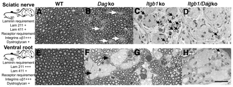
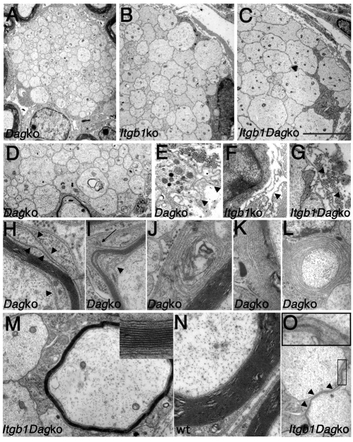
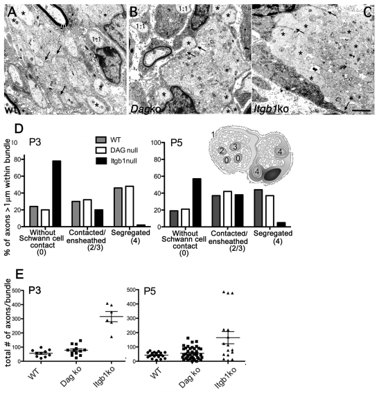
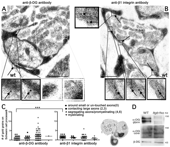
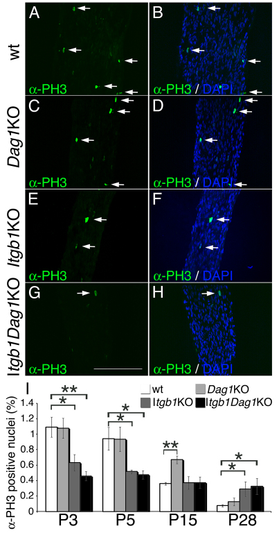
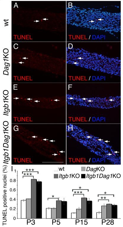
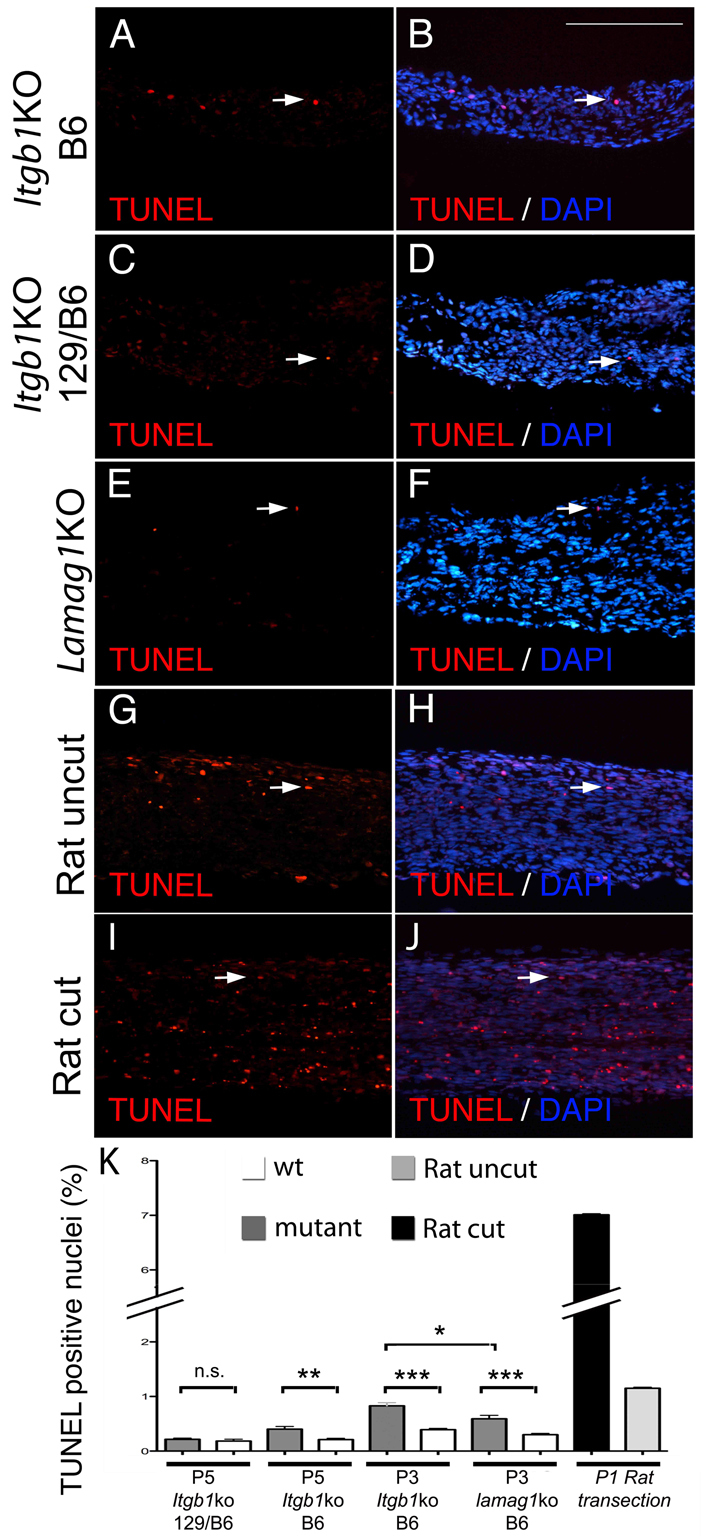
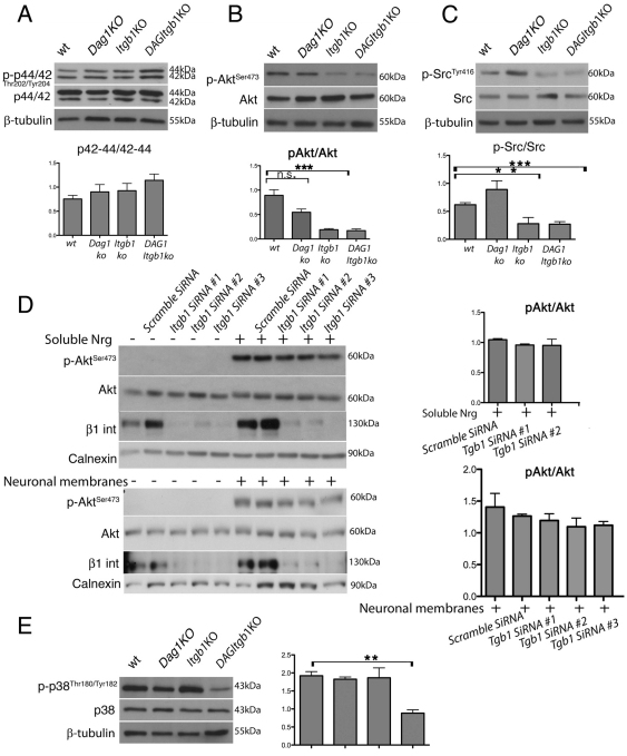
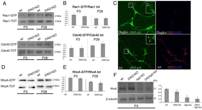
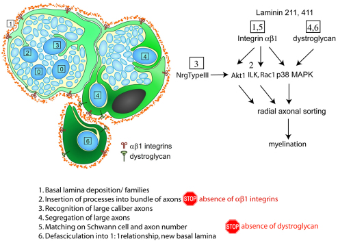
Radial sorting allows the segregation of axons by a single Schwann cell (SC) and is a prerequisite for myelination during peripheral nerve development. Radial sorting is impaired in models of human diseases, congenital muscular dystrophy (MDC) 1A, MDC1D and Fukuyama, owing to loss-of-function mutations in the genes coding for laminin α2, Large or fukutin glycosyltransferases, respectively. It is not clear which receptor(s) are activated by laminin 211, or glycosylated by Large and fukutin during sorting. Candidates are αβ1 integrins, because their absence phenocopies laminin and glycosyltransferase deficiency, but the topography of the phenotypes is different and β1 integrins are not substrates for Large and fukutin. By contrast, deletion of the Large and fukutin substrate dystroglycan does not result in radial sorting defects. Here, we show that absence of dystroglycan in a specific genetic background causes sorting defects with topography identical to that of laminin 211 mutants, and recapitulating the MDC1A, MDC1D and Fukuyama phenotypes. By epistasis studies in mice lacking one or both receptors in SCs, we show that only absence of β1 integrins impairs proliferation and survival, and arrests radial sorting at early stages, that β1 integrins and dystroglycan activate different pathways, and that the absence of both molecules is synergistic. Thus, the function of dystroglycan and β1 integrins is not redundant, but is sequential. These data identify dystroglycan as a functional laminin 211 receptor during axonal sorting and the key substrate relevant to the pathogenesis of glycosyltransferase congenital muscular dystrophies.


Figures

Fig. 1.
Fig. 1.
Axonal sorting requires both dystroglycan and β1 integrin in SCs. (A-H) Transverse semi-thin sections of sciatic nerves (A-D) and ventral roots (E-H) in dystroglycan (DagKO; B,F), β1 integrin (Itgβ1KO; C,G) and double SC-specific (D,H) null mice at P28. Laminins required in each district are indicated on the left (+, mild; +++, severe). In sciatic nerves, absence of dystroglycan (B) results in small bundles of unsorted axons (white arrows). The absence of β1 integrin (C) results in large bundles of unsorted axons (b) and few myelinated fibers (arrows). A complete arrest of radial sorting, with only an occasional myelinated fiber (arrow) is observed when both dystroglycan and β1 integrin are ablated (D). In ventral roots, the absence of either dystroglycan or β1 integrin causes an important arrest in radial sorting (F,G; arrows), which becomes complete when both receptors are ablated (H). dr, dorsal roots; sn, sciatic nerve; vr, ventral roots. Scale bar: 25 μm.
Fig. 2.
Fig. 2.
Radial sorting and myelin defects in dystroglycan and double mutants. Electron microscopy of transverse sections of dystroglycan, β1 integrin and double mutant sciatic nerves at P28. (A-D) Nerves of all the mutants contain bundles of naked axons (A-D), but the size of those axons is smaller in dystroglycan (A) compared with β1 integrin (B) and double mutants (C). (See also Table 2.) Occasionally, large axons are found in bundles of dystroglycan mutants (D). (E-G) Detached and redundant basal lamina (arrowheads) in β1 integrin (E), dystroglycan (F) and double (G) mutants. (H-L) Myelination defects in dystroglycan mutants: disorganization of inner and outer wraps (arrowheads in H,I) and outer mesaxons (arrow in I), non-compacted lamella in the SC cytoplasm (J), non-compaction of outer myelin lamellae (K) or throughout the whole myelin thickness (L). (M-O) Hypomyelination in double mutants (M). A myelin-like process was observed separating two axon bundles (arrowheads and inset in O). Wild type (wt) is shown in N. Scale bar: in C, 5 μm for A-C; 3.2 μm for D,O; 1.6 μm for E-G; 1.25 μm for M,N; 1 μm for J-L.
Fig. 3.
Fig. 3.
β1 integrins act before dystroglycan during axonal sorting. (A-C) Electron microscopy of cross-sections of P3 sciatic nerves of wt, dystroglycan and β1 integrin mutants. At this stage, wt nerves (A) contained SC families around small axonal bundles, with ∼45% of large axons already segregated at the periphery (asterisks), and pro-myelinating (1:1) and myelinating (m) fibers. SC processes enwrap axons (arrows). Radial sorting in dystroglycan mutants is delayed (B): SC families have bigger bundles, but ∼40% of large axons have been properly segregated to the periphery (asterisks) and pro-myelinating SC are frequent (1:1). In addition, SC start to enwrap axons with cytoplasmic processes (arrows). By contrast, β1 integrin mutant nerves (C) at this stage are completely occupied by large axonal bundles, and most large axons are trapped within bundles (asterisks). Cytoplasmic processes of SCs do not interact with axons (arrows). (D,E) Axon bundles in β1 integrin mutants are more immature than in dystroglycan mutants. (D) Quantification of the percentage of large axons in bundles based on their relationship with SCs, as indicated in the scheme: large axons with no association with SCs are stage 0, large axons contacted by SCs are stage 2 or 3, axons segregated at the periphery are stage 4. β1 integrin mutants have significantly more axons in stage 0 and fewer axons in stage 4 than do dystroglycan mutants (P<0.0001 by χ2 test, n=3 animals per genotype). (E) β1 integrin mutants have significantly more axons in bundles than do dystroglycan mutants (by Student's t-test: β1 integrin versus dystroglycan: P=0.0001 at P3 and 0.006 at P5; wt versus dystroglycan non significant, three animals/genotype). Mean ± s.e.m. are indicated.
Fig. 4
Fig. 4
. β-Dystroglycan is concentrated on SC processes segregating large axons. (A,B) Immuno-electron microscopy of P3-P4 mouse sciatic nerves using anti-β-dystroglycan (A) or anti-β1 integrin (B) antibodies shows that dystroglycan is detectable on SC processes surrounding large axons (arrows in A, magnified in the black insets; white insets show absence of dystroglycan on SC processes contacting unsegregated axons). By contrast, β1 integrins are detectable in all SCs surrounding all axons (arrows in B, magnified in the insets). Scale bar: 500 nm. (C) Number of gold grains found on SC processes at different stages (0 to 6 as shown in the drawing and in Fig. 10). More gold grains are present on immature SC processes (stage 0) when using the anti-β1 integrin antibody than when using anti-β-dystroglycan antibody (P=0.004 by Student's t-test, n=35 processes for β-dystroglycan, 39 for β1 integrin from at least three different experiments). The number of gold grains marking β-dystroglycan significantly increases in SCs segregating large caliber axons and pro-myelinating SCs (stages 4,6). Mean ± s.e.m. are indicated. See also Table 3. (D) Western blot from P28 sciatic nerves shows that the α-chain of dystroglycan (DG) is hypoglycosylated in the absence of β1-integrins.
Fig. 5.
Fig. 5.
SC proliferation is decreased in β1-integrin mutants. (A-H) Immunostaining with anti-phospho-histone H3 (green) and staining with the nuclear dye DAPI (blue in the merged image in B,D,F,H) on longitudinal sections of control (A,B) and mutant (C-H) mice (P3 sciatic nerves) to measure the fraction of proliferating nuclei (arrows). Scale bar: 100 μm. (I) Quantification shows that the rate of proliferation is decreased in β1 integrin and double mutants at P3 and P5, but is normal by P15 and is increased later. By contrast, SC lacking dystroglycan do not show decreased proliferation. Error bars indicate s.e.m. *P≤0.05, **P≤0.005 by Student's t-test, n=3 mice per genotype.
Fig. 6.
Fig. 6.
SC survival is impaired in newborn β1 integrin mutants. (A-H) TUNEL analysis (red) on longitudinal sections of control (A,B) and mutant (C-H) mice (P3 sciatic nerves) and staining with the nuclear dye DAPI (blue in the merged image in B,D,F,H). Arrows indicate TUNEL-positive nuclei. Scale bar: 100 μm. (I) Apoptosis is increased in β1 integrin and double mutants. Error bars indicate s.e.m. *P≤0.05, **P≤0.005, ***P≤0.0005, by Student's t-test, n=3 mice per genotype.
Fig. 7.
Fig. 7.
The moderate increase in apoptosis in SCs lacking β1 integrins is comparable to that observed in the absence of all laminins. (A-K) TUNEL on longitudinal sections of mutant mice at P5 (A-D), P3 (E,F) or P1 rat wt uncut and cut (G-J) sciatic nerves. (C,D,K) TUNEL on P5 β1 integrin mutant in a mixed C57BL6/129sv background (N2–N5 C57BL6) shows no difference between mutant and wt nerves, in agreement with previous data (Feltri et al., 2002). (A,B,K) By contrast, the mice used in this study, >N15 congenic in C57BL6, show a small but significant increase in apoptosis (see also Fig. 6). (E,F,K). Comparable levels of apoptosis are detected in laminin γ1 mutants (laminin γ1 KO). The increase in apoptosis (7%) detected after transection of rat sciatic nerve at P1 is significantly higher, as reported previously (Grinspan et al., 1996). *P<0.01, **P<0.001, ***P<0.0001, n.s., non significant by Student's t-test, minimum three mice per genotype. Error bars indicate s.e.m. Scale bar: 100 μm.
Fig. 8.
Fig. 8.
Impaired Akt, Src and p38MAPK activation during sorting. (A-C,E) Western blots of P3 sciatic nerves pooled from three to six animals from the indicated genotypes. Graphs indicate mean of at least three experiments using different pools of animals. Error bars indicate s.e.m. *P≤0.05, **P≤0.005, ***P≤0.0005, n.s., non significant, by Student's t-test. (B) Western blot using anti-p-AktSer473 antibodies and normalized for total Akt levels show a significant decrease in p-AktSer473 only in β1 mutants. (A) p-P44/42 levels are similar in mutant and wt. (C) p-SrcTyr416 is decreased in β1 integrin mutants whereas p-p38Thr180/Tyr182 (E) is decreased only when both β1 integrins and dystroglycan are deleted. (E) Western blot shows the efficient silencing of β1 integrin, and the appropriate phosphorylation of Akt on Ser473 in β1 integrin-silenced SCs. (D) Rat SCs transfected with either β1 integrin silencing or control siRNA, starved and stimulated with either the soluble EGF domain or dorsal root ganglia membranes containing neuregulin-type III (Taveggia et al., 2005).
Fig. 9.
Fig. 9.
Normal activation of small Rho GTPases in the absence of SC dystroglycan. (A,B,D,E) The GTP-bound fraction of small GTPases in P3 and P28 sciatic nerve were measured using pull-down assays. Active proteins were normalized to total Rac1, Cdc42 and RhoA. (F) Western blot for RhoE in P3 sciatic nerves. (C) SCs plated on laminin, treated with GST-PBD and saponin, and stained with anti-GST (green) and anti-dystroglycan (red) antibodies show that in the absence of dystroglycan, active Rac1 is correctly targeted to lamellipodia. The leading edge of lamellipodia is enriched with PBD (Pak binding domain) in both wt and dystroglycan-null cells (arrowheads in the enlarged insets). Graphs represent the mean of at least three different experiments, error bars indicate s.e.m.
Fig. 10.
Fig. 10.
Radial sorting is a multistep process. Schematic of the morphogenetic steps showing that deposition of the basal lamina and formation of `families' (step 1) requires laminins, insertion of processes into bundles (step 2) requires αβ1 integrins, whereas defasciculation of axons into pro-myelinating SCs with their own basal lamina (steps 4-6) requires dystroglycan (this paper). The presumed involvement of other signals (Nrg1 type III, Akt, Ilk, Rac1 and p38 MAPK) at various steps is also indicated.

References

    1. Barresi R., Campbell K. P. (2006). Dystroglycan: from biosynthesis to pathogenesis of human disease. J. Cell Sci. 119, 199-207 - PubMed
    1. Benninger Y., Thurnherr T., Pereira J. A., Krause S., Wu X., Chrostek-Grashoff A., Herzog D., Nave K. A., Franklin R. J., Meijer D., et al. (2007). Essential and distinct roles for cdc42 and rac1 in the regulation of Schwann cell biology during peripheral nervous system development. J. Cell Biol. 177, 1051-1061 - PMC - PubMed
    1. Bhat N. R., Zhang P., Mohanty S. B. (2007). p38 MAP kinase regulation of oligodendrocyte differentiation with CREB as a potential target. Neurochem.Res. 32, 293-302 - PubMed
    1. Bradley W. G., Jenkison M. (1973). Abnormalities of peripheral nerves in murine muscular dystrophy. J. Neurol. Sci. 18, 227-247 - PubMed
    1. Bradley W. G., Jenkison M. (1975). Neural abnormalities in the dystrophic mouse. J. Neurol. Sci. 25, 249-255 - PubMed

Publication types

MeSH terms

Supplementary concepts