

Regulation of tooth number by fine-tuning levels of receptor-tyrosine kinase signaling
- PMID: 21862563
- PMCID: PMC3160100
- DOI: 10.1242/dev.069195
Regulation of tooth number by fine-tuning levels of receptor-tyrosine kinase signaling
Abstract
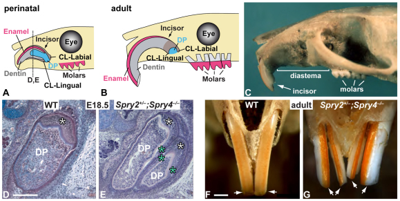
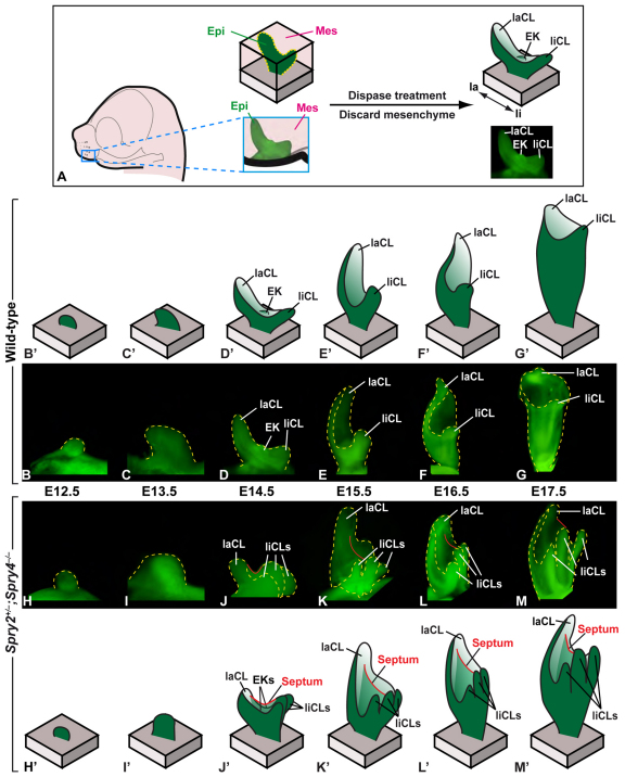
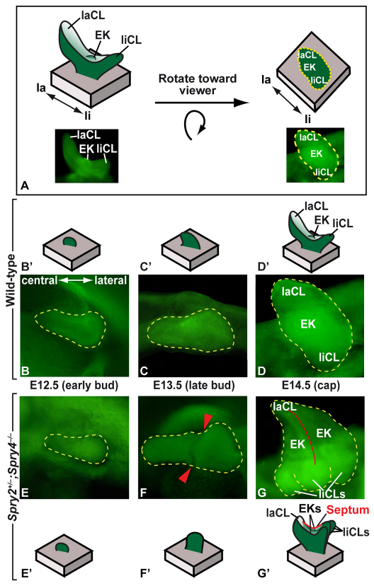
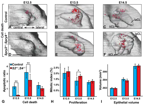
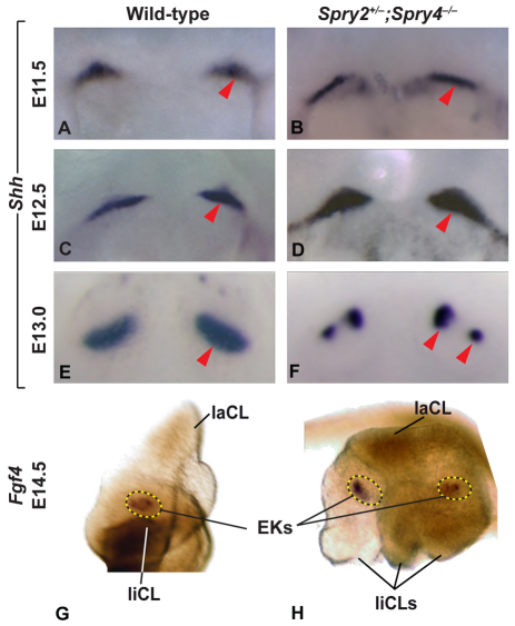
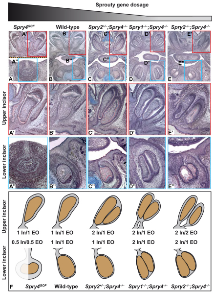
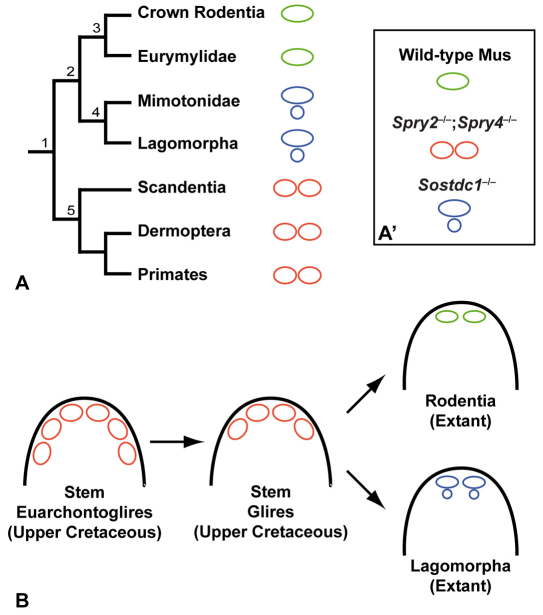
Much of our knowledge about mammalian evolution comes from examination of dental fossils, because the highly calcified enamel that covers teeth causes them to be among the best-preserved organs. As mammals entered new ecological niches, many changes in tooth number occurred, presumably as adaptations to new diets. For example, in contrast to humans, who have two incisors in each dental quadrant, rodents only have one incisor per quadrant. The rodent incisor, because of its unusual morphogenesis and remarkable stem cell-based continuous growth, presents a quandary for evolutionary biologists, as its origin in the fossil record is difficult to trace, and the genetic regulation of incisor number remains a largely open question. Here, we studied a series of mice carrying mutations in sprouty genes, the protein products of which are antagonists of receptor-tyrosine kinase signaling. In sprouty loss-of-function mutants, splitting of gene expression domains and reduced apoptosis was associated with subdivision of the incisor primordium and a multiplication of its stem cell-containing regions. Interestingly, changes in sprouty gene dosage led to a graded change in incisor number, with progressive decreases in sprouty dosage leading to increasing numbers of teeth. Moreover, the independent development of two incisors in mutants with large decreases in sprouty dosage mimicked the likely condition of rodent ancestors. Together, our findings indicate that altering genetic dosage of an antagonist can recapitulate ancestral dental characters, and that tooth number can be progressively regulated by changing levels of activity of a single signal transduction pathway.
Figures
Similar articles
-
The dynamics of supernumerary tooth development are differentially regulated by Sprouty genes.J Exp Zool B Mol Dev Evol. 2013 Jul;320(5):307-20. doi: 10.1002/jez.b.22502. Epub 2013 Apr 19. J Exp Zool B Mol Dev Evol. 2013. PMID: 23606267
-
An FGF signaling loop sustains the generation of differentiated progeny from stem cells in mouse incisors.Development. 2008 Jan;135(2):377-85. doi: 10.1242/dev.015081. Epub 2007 Dec 12. Development. 2008. PMID: 18077585 Free PMC article.
-
Sprouty genes control diastema tooth development via bidirectional antagonism of epithelial-mesenchymal FGF signaling.Dev Cell. 2006 Aug;11(2):181-90. doi: 10.1016/j.devcel.2006.05.014. Dev Cell. 2006. PMID: 16890158 Free PMC article.
-
Sprouty proteins, masterminds of receptor tyrosine kinase signaling.Angiogenesis. 2008;11(1):53-62. doi: 10.1007/s10456-008-9089-1. Epub 2008 Jan 25. Angiogenesis. 2008. PMID: 18219583 Review.
-
Development of the vestigial tooth primordia as part of mouse odontogenesis.Connect Tissue Res. 2002;43(2-3):120-8. doi: 10.1080/03008200290000745. Connect Tissue Res. 2002. PMID: 12489147 Review.
Cited by
-
Pax9's Interaction With the Ectodysplasin Signaling Pathway During the Patterning of Dentition.Front Physiol. 2020 Nov 26;11:581843. doi: 10.3389/fphys.2020.581843. eCollection 2020. Front Physiol. 2020. PMID: 33329029 Free PMC article.
-
Sprouty/FGF signaling regulates the proximal-distal feather morphology and the size of dermal papillae.Dev Biol. 2012 Dec 1;372(1):45-54. doi: 10.1016/j.ydbio.2012.09.004. Epub 2012 Sep 18. Dev Biol. 2012. PMID: 23000358 Free PMC article.
-
Notum regulates the cusp and root patterns in mouse molar.Sci Rep. 2024 Jun 13;14(1):13633. doi: 10.1038/s41598-024-64340-w. Sci Rep. 2024. PMID: 38871845 Free PMC article.
-
Ontogenetic variations and structural adjustments in mammals evolving prolonged to continuous dental growth.R Soc Open Sci. 2017 Jul 26;4(7):170494. doi: 10.1098/rsos.170494. eCollection 2017 Jul. R Soc Open Sci. 2017. PMID: 28791172 Free PMC article.
-
Common developmental pathways link tooth shape to regeneration.Dev Biol. 2013 May 15;377(2):399-414. doi: 10.1016/j.ydbio.2013.02.007. Epub 2013 Feb 17. Dev Biol. 2013. PMID: 23422830 Free PMC article.
References
-
- Asher R., Meng J., Wible J., McKenna M., Rougier G., Dashzeveg D., Novacek M. (2005). Stem lagomorpha and the antiquity of Glires. Science 307, 1091-1094 - PubMed
-
- Basson M., Akbulut S., Watson-Johnson J., Simon R., Carroll T., Shakya R., Gross I., Martin G., Lufkin T., McMahon A., et al. (2005). Sprouty1 is a critical regulator of GDNF/RET-mediated kidney induction. Dev. Cell 8, 229-239 - PubMed
Publication types
MeSH terms
Substances
Grants and funding
LinkOut - more resources
Full Text Sources
Molecular Biology Databases
Research Materials
