. 2011 Oct 14;286(41):35663-35674.
doi: 10.1074/jbc.M111.267567. Epub 2011 Aug 23.

Polyubiquitin binding to optineurin is required for optimal activation of TANK-binding kinase 1 and production of interferon β

Affiliations

Polyubiquitin binding to optineurin is required for optimal activation of TANK-binding kinase 1 and production of interferon β

Catherine E Gleason et al. J Biol Chem. .

Abstract

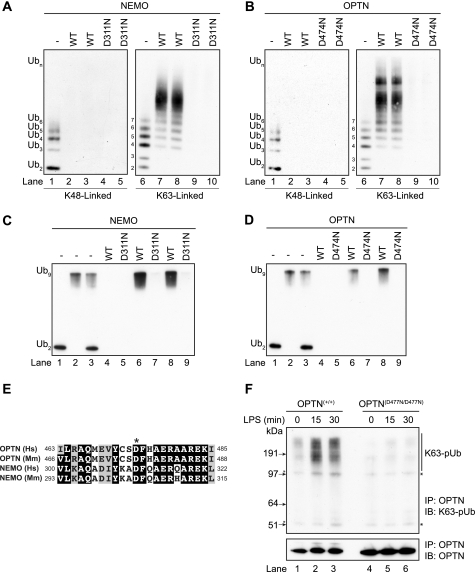
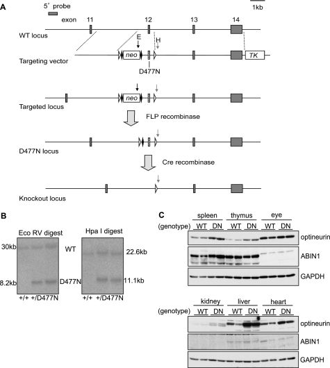
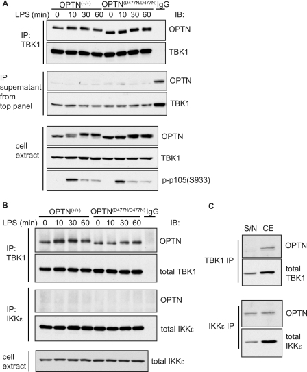
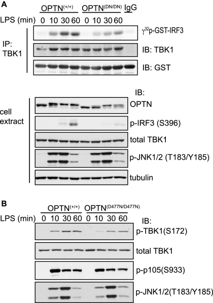
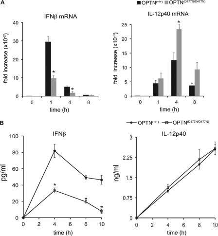
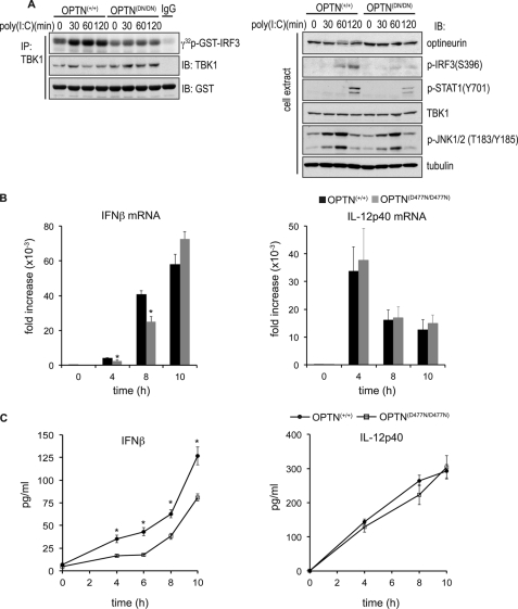
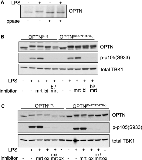
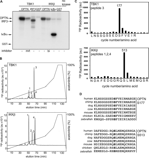
TANK-binding kinase (TBK1) is essential for transcription of the interferon (IFN) β gene in response to lipopolysaccharide (LPS) and double-stranded RNA, but the molecular mechanisms that underlie the activation of TBK1 are incompletely understood. Previously, we identified the NF-κB essential modulator (NEMO)-related polyubiquitin-binding protein, optineurin (OPTN), as a novel binding partner of TBK1. To determine whether the ubiquitin-binding function of OPTN is involved in regulating TBK1 and IFNβ production, we generated a mouse in which wild-type optineurin was replaced by the polyubiquitin binding-defective mutant, OPTN(D477N/D477N). In this study, we found that LPS or poly(I:C)-induced TBK1 activity was significantly reduced in bone marrow-derived macrophage (BMDM) from OPTN(D477N/D477N) mice. Consistent with this, the phosphorylation of IFN regulatory factor 3 (IRF3) and the production of IFNβ mRNA and secretion were reduced. Stimulation of BMDMs with LPS triggered the phosphorylation of OPTN, which was reversed by phosphatase treatment and prevented by pharmacological inhibition of both the canonical IκB kinases (IKKα/β) and the IKK-related kinases (TBK1/IKKε). In contrast, LPS-stimulated phosphorylation of OPTN(D477N) was markedly reduced in BMDMs from OPTN(D477N/D477N) mice, and inhibition of the canonical IKKs alone prevented phosphorylation, providing further evidence that ubiquitin binding to OPTN contributes to LPS-induced TBK1 activation. TBK1 and IKKβ phosphorylated OPTN preferentially at Ser-177 and Ser-513, respectively, in vitro. In conclusion, our results suggest that OPTN binds to polyubiquitylated species formed in response to LPS and poly(I:C), enhancing the activation of TBK1 that is required for optimal phosphorylation of IRF3 and production of IFNβ.


Figures

FIGURE 1.
FIGURE 1.
Optineurin binds to Lys-63-linked and linear polyubiquitin chains. A and B, method used to study the binding of polyubiquitin chains has been described previously for GST-NEMO (17). Polyubiquitin chains captured by the immobilized human GST-NEMO (A) and human GST-OPTN (B) were released by denaturation in 1% (w/v) SDS, subjected to SDS-PAGE, and immunoblotted with an anti-ubiquitin antibody. Lanes 1 and 6 show, respectively, the Lys-48- and Lys-63-linked polyubiquitin preparations used in the experiment. The Lys-48-linked (lanes 2 and 3) and Lys-63-linked (lanes 7 and 8) polyubiquitin chains captured by NEMO (left-hand panel) or OPTN (right-hand panel) are shown. The OPTN(D474N) and NEMO(D311N) mutants did not bind to either Lys-48- or Lys-63-linked polyubiquitin chains under the conditions used (lanes 4, 5, 9, and 10 in the left- and right-hand panels). C and D, as in A and B except that binding to linear polyubiquitin oligomers was studied. Lanes 1–3 show the di-ubiquitin and nona-ubiquitin preparations used in the experiment. Lanes 4, 6, and 8 show that NEMO (C) and OPTN (D) bind to nona-ubiquitin but not to di-ubiquitin under the conditions used. Lanes 5, 7, and 9 show that NEMO(D311N) and OPTN(D474N) do not bind to linear polyubiquitin oligomers. Similar results were obtained in two different experiments. E, amino acid sequences in NEMO and OPTN surrounding the aspartic acid residue (*) in the UBAN that is critical for binding to polyubiquitin chains. Identities are shown by the white lettering on a black background and similarities by the black letters on a gray background. Abbreviations used are as follows: Hs, Homo sapiens; Mm, Mus musculus. F, BMDMs from OPTN+/+ or OPTND477N/D477N mice were stimulated with LPS (100 ng/ml) for the times indicated and extracted in lysis buffer containing 100 mm iodoacetamide to inhibit deubiquitylases. OPTN was then immunoprecipitated (IP) from 10 mg of cell extract protein using 40 μg of anti-human OPTN coupled covalently to protein-G-Sepharose. After 3 h at 4 °C, the immunoprecipitates were washed five times with lysis buffer plus 500 mm NaCl and once with 10 mm Tris/HCl, pH 8.0, before denaturation in SDS followed by SDS-PAGE and immunoblotting (IB) with antibodies that recognize K63-pUb chains or OPTN. The asterisks denote nonspecific bands.
FIGURE 2.
FIGURE 2.
Generation of OPTND477N/D477N knock-in mice. A, strategy for generating the D477N knock-in mutation in the mouse optn gene. A vector was used to introduce a point mutation producing the D477N mutation into exon 12 of the OPTN locus and loxP sites (open triangles) either side of exon 12. The vector also contained a neomycin resistance cassette (neo) flanked by FLP recognition target sites (closed diamonds) for positive selection and a thymidine kinase gene (TK) for negative selection. The vector was used to target ES cells, which were screened by Southern blots using a probe 5′ to the targeting vector. The vector introduced an additional EcoRV site (black arrow, E) in the neo gene and an HpaI site (gray arrow, H) 3′ to the second loxP site. B, using the 5′ probe, EcoRV and HpaI digests gave bands of 30 or 22.6 kb, respectively, for a wild-type allele or 8.2 and 11.1 kb for a targeted locus. ES cells were used to generate knock-in mice from which the neo gene was removed by crossing to FLPe transgenic mice. C, comparison of the expression of the optineurin and ABIN1 proteins in various mouse tissues from OPTN+/+ (WT) or OPTND477N/D477N mice (DN) as judged by immunoblotting. Each lane represents tissue lysate (50 μg of protein) from an individual mouse (n = 2 per genotype). Glyceraldehyde 3-phosphate dehydrogenase (GAPDH) was used as the loading control.
FIGURE 3.
FIGURE 3.
Optineurin-TBK1 interaction does not depend on polyubiquitin binding and occurs independently of stimulation. BMDMs from OPTN+/+ and OPTND477N/D477N mice were stimulated with 100 ng/ml LPS for 10, 30, or 60 min or left unstimulated (0 min). A, top two panels, TBK1 was immunoprecipitated (IP) from the cell lysates using anti-TBK1 or control IgG, and the presence of OPTN and TBK1 in the immunoprecipitates was analyzed by immunoblotting (IB). A, middle two panels, supernatants obtained after immunoprecipitation of TBK1 were also analyzed by immunoblotting to assess the extent of depletion of OPTN and TBK1 from the cell extracts. A, bottom panels, cell lysates were immunoblotted with the indicated antibodies. B, TBK1 or IKKϵ was immunoprecipitated (IP) from 50 μg of cell extract protein using anti-TBK1, anti-IKKϵ, or control IgG, and the presence of OPTN, TBK1, or IKKϵ in the immunoprecipitates was analyzed by immunoblotting (IB). Cell extracts (30 μg) were immunoblotted to measure total levels of IKKϵ. C, amount of OPTN and TBK1 remaining in the supernatant (S/N) and cell extract (CE) obtained after immunoprecipitation of TBK1 (top two panels) or IKKϵ (bottom two panels) from the unstimulated OPTN+/+ samples. Results for A–C are representative of at least three separate experiments.
FIGURE 4.
FIGURE 4.
Polyubiquitin binding to optineurin is required for full TBK1 activation in response to LPS. OPTN+/+ and OPTND477N/D477N BMDMs were stimulated with 100 ng/ml LPS for the times indicated. A, in the upper panel, TBK1 was immunoprecipitated (IP) from 50 μg of cell extract protein and assayed with GST-IRF3 and Mg-[γ-32P]ATP as the substrates (see “Experimental Procedures”). The reactions were stopped by denaturation in SDS, subjected to SDS-PAGE, and after transfer to a PVDF membrane were autoradiographed to detected the 32P-labeled GST-IRF3 formed during the assay. The total levels of TBK1 and GST-IRF3 present in each assay were assessed by immunoblotting (IB). In the lower panel, cell extract from each experiment (30 μg of protein) was analyzed by immunoblotting for the total levels of OPTN, TBK1, and tubulin and for phosphorylated IRF3 and phosphorylated JNK1/2. B, experiment was carried out as in A, except that the cell extracts were immunoblotted for TBK1 phosphorylated at Ser-172 as well as for total TBK1 and the phosphorylation of p105 and JNK. A and B, results are representative of at least three separate experiments.
FIGURE 5.
FIGURE 5.
LPS-stimulated production of IFNβ is reduced in BMDMs from OPTND477N/D477N mice. BMDMs from OPTN+/+ and OPTND477N/D477N mice were stimulated with 100 ng/ml LPS for the times indicated. A, total RNA was extracted and analyzed by quantitative PCR for expression of IFNβ (left-hand panel) and IL-12p40 mRNA levels (right-hand panel). mRNA levels were normalized for 18 S rRNA expression, and the results are presented as fold-increase relative to the mRNA levels present in wild-type unstimulated cells. B, concentration of IFNβ (left-hand panel) or IL-12p40 (right-hand panel) present in the cell culture medium as measured by ELISA. Results are representative of at least two separate experiments with n = 4 mice per genotype. The error bars represent the mean ± S.E. Statistical analysis was performed by one-way analysis of variance with p < 0.05 considered statistically significant (*).
FIGURE 6.
FIGURE 6.
Poly(I:C)-induced TBK1 activity and IFNβ production are reduced in OPTND477N/D477N BMDMs. OPTN+/+and OPTND477N/D477N BMDMs were stimulated with 10 μg/ml poly(I:C) for the times indicated. A, TBK1 activities (left-hand panel) measured as described in Fig. 4. Further aliquots of the extracts (30 μg of protein) were immunoblotted with the same antibodies used in Fig. 4A, as well as for STAT1 phosphorylated at Tyr-701. B, total RNA was extracted and analyzed by quantitative RT-PCR for expression of IFNβ and IL-12p40 mRNA levels. The mRNA levels were normalized for 18 S rRNA expression, and the results are presented as the fold-increase from wild-type unstimulated cells. C, concentrations of IFNβ or IL-12p40 in the culture media were determined by ELISA. Results are representative of at least three separate experiments with n = 4 mice per genotype. The error bars represent the mean ± S.E. Statistical analysis was performed by one-way analysis of variance with p < 0.05 considered statistically significant (*).
FIGURE 7.
FIGURE 7.
Optineurin is phosphorylated in response to LPS in a TBK1- and TAK1-IKKα/β-dependent manner. BMDMs from OPTN+/+ and OPTND477N/D477N mice were stimulated for 30 min with 100 ng/ml LPS. A, wild-type optineurin was immunoprecipitated from 50 μg of cell extract protein of OPTN+/+ BMDMs using a sheep α-mouse optineurin antibody. The resuspended immunoprecipitates (30 μl) were then treated with (+) or without (−) the protein phosphatase from bacteriophage λgt10 (λ-ppase) (200 units). The change in mobility of OPTN following treatment with λppase was analyzed by immunoblotting with an anti-OPTN antibody. B, prior to stimulation with LPS, BMDMs from OPTN+/+ and OPTND477N/D477N mice were incubated for 1 h with or without (−) the TBK1/IKKϵ inhibitor MRT67307 (mrt), the IKKβ inhibitor BI605906 (bi), or both inhibitors. The cell extracts were then subjected to SDS-PAGE followed by immunoblotting with the antibodies indicated. The inhibition of IKKβ was confirmed by immunoblotting for phosphorylated p105 (S933). C, same as B, except that the TAK1 inhibitor 5Z-7-oxozeanol (ox) was used instead of BI605906 to inhibit the activation of both IKKα and IKKβ. The inhibition of IKKβ or TAK1 was confirmed by immunoblotting for phosphorylated p105 (S933). A–C, results are representative of at least two separate experiments.
FIGURE 8.
FIGURE 8.
Identification of sites on optineurin phosphorylated by TBK1 or IKKβ in vitro. A, wild-type GST-OPTN (2 μm) was incubated for 30 min with TBK1 (2 units/ml) or IKKβ (2 units/ml) in the absence (−) or presence of MRT67307 (mrt) or BI605906 (bi) prior to initiating the protein kinase reactions with Mg-[γ-32p]ATP. The phosphorylation of OPTN was compared with the phosphorylation of GST-IRF3 or GST-IκBα(2–54) (2 μm), which are established physiological substrates of TBK1 and IKKβ, respectively. GST (2 μm) was used as control. The reactions were terminated in SDS and subjected to SDS-PAGE, and the gel was autoradiographed. B, 32P-labeled OPTN from A obtained by phosphorylation with TBK1 (upper panel) or IKKβ (lower panel) were digested with trypsin and subjected to chromatography on a Vydac C18 column as described under “Experimental Procedures.” 32P radioactivity in arbitrary units (au) is shown by the full line, and the acetonitrile gradient is indicated by the diagonal lines. C, solid phase sequencing of peptide 3 generated after phosphorylation by TBK1 (upper panel of B) and the pooled peptides 1, 2, and 4 generated after phosphorylation by IKKβ (lower panel of B). Peptides were subjected to solid phase sequencing (49) to identify the cycles of Edman degradation at which 32P radioactivity (filled bars) was released from the phosphopeptides present in these fractions. D, alignment of amino acid sequences of OPTN from various species demonstrating conservation of the sites phosphorylated by TBK1 or IKKβ in vertebrate species. The phosphorylated serine residues are shown in boldface type.

Similar articles

Cited by

References

    1. Walsh M. C., Kim G. K., Maurizio P. L., Molnar E. E., Choi Y. (2008) PLoS One 3, e4064. - PMC - PubMed
    1. Wang C., Deng L., Hong M., Akkaraju G. R., Inoue J., Chen Z. J. (2001) Nature 412, 346–351 - PubMed
    1. Haas T. L., Emmerich C. H., Gerlach B., Schmukle A. C., Cordier S. M., Rieser E., Feltham R., Vince J., Warnken U., Wenger T., Koschny R., Komander D., Silke J., Walczak H. (2009) Mol. Cell 36, 831–844 - PubMed
    1. Kirisako T., Kamei K., Murata S., Kato M., Fukumoto H., Kanie M., Sano S., Tokunaga F., Tanaka K., Iwai K. (2006) EMBO J. 25, 4877–4887 - PMC - PubMed
    1. Tokunaga F., Sakata S., Saeki Y., Satomi Y., Kirisako T., Kamei K., Nakagawa T., Kato M., Murata S., Yamaoka S., Yamamoto M., Akira S., Takao T., Tanaka K., Iwai K. (2009) Nat. Cell Biol. 11, 123–132 - PubMed

Publication types

MeSH terms