. 2011 Oct 7;286(40):34923-31.
doi: 10.1074/jbc.M111.239350. Epub 2011 Aug 23.

A molecular chaperone mediates a two-protein enzyme complex and glycosylation of serine-rich streptococcal adhesins

Affiliations

A molecular chaperone mediates a two-protein enzyme complex and glycosylation of serine-rich streptococcal adhesins

Ren Wu et al. J Biol Chem. .

Abstract

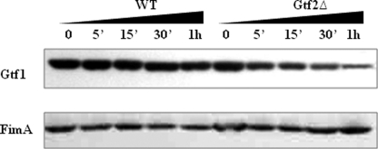
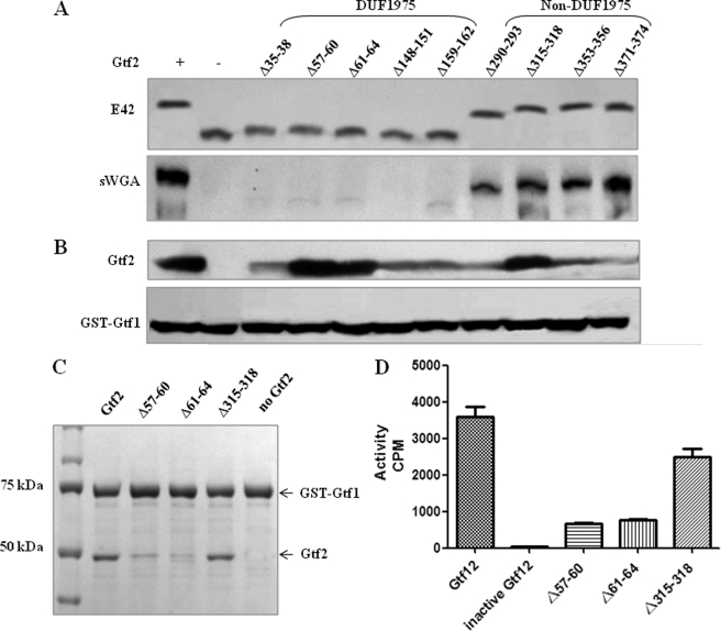
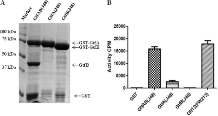
Serine-rich repeat glycoproteins identified from streptococci and staphylococci are important for bacterial adhesion and biofilm formation. Two putative glycosyltransferases, Gtf1 and Gtf2, from Streptococcus parasanguinis form a two-protein enzyme complex that is required for glycosylation of a serine-rich repeat adhesin, Fap1. Gtf1 is a glycosyltransferase; however, the function of Gtf2 is unknown. Here, we demonstrate that Gtf2 enhances the enzymatic activity of Gtf1 by its chaperone-like property. Gtf2 interacted with Gtf1, mediated the subcellular localization of Gtf1, and stabilized Gtf1. Deletion of invariable amino acid residues in a conserved domain of unknown function (DUF1975) at the N terminus of Gtf2 had a greater impact on Fap1 glycosylation than deletion of the C-terminal non-DUF1975 residues. The DUF1975 deletions concurrently reduced the interaction between Gtf1 and Gtf2, altered the subcellular localization of Gtf1, and destabilized Gtf1, suggesting that DUF1975 is crucial for the chaperone activity of Gtf2. Homologous GtfA and GtfB from Streptococcus agalactiae rescued the glycosylation defect in the gtf1gtf2 mutant; like Gtf2, GtfB also possesses chaperone-like activity. Taken together, our studies suggest that Gtf2 and its homologs possess the conserved molecular chaperone activity that mediates protein glycosylation of bacterial adhesins.


Figures

FIGURE 1.
FIGURE 1.
Gtf2 stabilizes Gtf1. S. parasanguinis cells were treated with 300 μg/ml chloramphenicol to stop nascent protein synthesis. 0.5 ml of cells was harvested 5, 15, and 30 min and 1 h post-treatment. The harvested bacteria were separated by SDS-PAGE. The stability of endogenous Gtf1 in the wild type and the gtf2 mutant was analyzed by Western blotting using anti-Gtf1 mAb. FimA was probed and used as a loading control.
FIGURE 2.
FIGURE 2.
Gtf2 modulates the subcellular distribution of Gtf1, and Gtf1 localized to the membrane fraction is resistant to trypsin digestion. A, distribution of Gtf1 in cell wall-free (lanes 1, 4, and 7), cytoplasmic (lanes 2, 5, and 8), and membrane (lanes 3, 6, and 9) fractions in the wild type (lanes 1–3), the gtf2 mutant (lanes 4–6), and the complemented strain (lanes 7–9). B, distribution of Gtf1 in cell wall-free (lanes 1, 4, and 7), cytoplasmic (lanes 2, 5, and 8), and membrane (lanes 3, 6, and 9) fractions in wild-type (lanes 1–3), the gap1 mutant (lanes 4–6), and the secY2 mutant (lanes 7–9). The subcellular localization of Gtf1 was analyzed by Western blotting using anti-Gtf1 mAb. The membrane-associated protein FimA was used as a loading and membrane fraction control. C, cytoplasmic and membrane fractions prepared from S. parasanguinis were subjected to trypsin digestion and analyzed by Western blotting using anti-Gtf1 mAb for Gtf1 stability.
FIGURE 3.
FIGURE 3.
DUF1975 modulates Fap1 glycosylation. Recombinant E. coli strains that harbor mini Fap1 (Fap1ΔRII) were transformed with pGEX6p1-gtf1-gtf2 or constructs encoding Gtf1 and different Gtf2 variants and characterized. A, effect of DUF1975 on Fap1 production. Cell lysates from wild-type Gtf2 and Gtf2 variants were probed with mAb E42 for Fap1 production (upper panel) and with succinyl wheat germ agglutinin (sWGA) for GlcNAc modification (lower panel). B, effect of DUF1975 on production of recombinant Gtf2 and Gtf1. Cell lysates from wild-type Gtf2 and Gtf2 variants were probed with anti-GST antibody for recombinant Gtf1 production (lower panel) and with anti-Gtf2 mAb for Gtf2 production (upper panel). C, effect of DUF1975 on Gtf1-Gtf2 interaction. His-tagged Gtf2 variants were purified and dissolved in NETN (20 mm Tris-HCl, 0.1 m NaCl, 1 mm EDTA, 0.5% NP-40, pH 7.2) buffer. The same amount of purified Gtf2 variants was incubated with GST-Gtf1 immobilized on glutathione beads to carry out GST pulldown assays. The Gtf2 variants bound to the beads were eluted and subjected to SDS-PAGE analysis and Coomassie Blue staining and used to measure the strength of the protein-protein interaction. D, effect of DUF1975 on enzymatic activity. The same amount of Gtf1-Gtf2 complexes purified from wild-type Gtf2 or the Gtf2 variants was used in an in vitro glycosyltransferase assay as described under “Experimental Procedures” to determine enzymatic activity. Samples were prepared and analyzed in triplicate. Error bars represent S.D.
FIGURE 4.
FIGURE 4.
DUF1975 modulates the stability of Gtf1 and Fap1. The stability of Gtf1 and Fap1 coexpressed with Gtf2 variants (wild-type Gtf2, Gtf2Δ, Δ57–60, and Δ315–318) in a recombinant E. coli glycosylation system was determined by inhibiting nascent protein synthesis. Cells that carry wild-type Gtf2 and Gtf2 variants were treated with tetracycline at 30 μg/ml. 0.5 ml of cell culture was collected 15 and 30 min and 1 h post-treatment. The same number of harvested bacterial cells was analyzed by Western blotting using antibodies recognizing recombinant Gtf2, Gtf1, and Fap1, respectively.
FIGURE 5.
FIGURE 5.
Gtf2 protects Gtf1 from proteolytic degradation by trypsin. Different amounts (0.04 ng to 4 μg) of Gtf2 were mixed with Gtf1 (4 μg in 20 mm Tris-HCl and 0.1 m NaCl, pH 8.0) (a 25-kDa putative galactosyltransferase 2 (GalT2) domain protein was used to compensate for Gtf2 missing in each sample) and then subjected to trypsin digestion. After the digest, the samples were subjected to SDS-PAGE analysis, and the Gtf1 proteins that remained in each sample were visualized by Coomassie Blue staining.
FIGURE 6.
FIGURE 6.
DUF1975 modulates the subcellular distribution of Gtf1 and the proteolytic resistance of Gtf1. A and B, the subcellular distribution of endogenous Gtf1 in cell wall-free (lanes 1, 4, 7, 10, and 13), cytoplasmic (lanes 2, 5, 8, 11, and 14), and membrane (lanes 3, 6, 9, 12, and 15) fractions in wild-type Gtf2 (A, lanes 1–3) and different Gtf2 variants (A, lanes 4–15, and B, lanes 1–6) was analyzed by Western blotting using anti-Gtf1 mAb. C, proteolytic resistance of Gtf1 in whole cell lysates. The same amount of whole cell lysates prepared from S. parasanguinis bacterial cells was digested by 4 μg/ml trypsin in the presence or absence of 0.5% Triton X-100 at 37 °C. D, proteolytic resistance of Gtf1 in membrane fractions. Membrane fractions prepared from the Δ57–60 and Δ315–318 variants were subjected to trypsin digestion as described above. The presence of Gtf1 was determined by Western blotting using anti-Gtf1 mAb. The membrane-associated protein FimA was used as a loading and fraction control.
FIGURE 7.
FIGURE 7.
The Gtf complex from S. agalactiae exhibits glycosyltransferase activity. A, co-purification of GtfA with GtfB. Recombinant E. coli strains that carry three constructs that express GST-GtfA/B, GST-GtfA, and GST-GtfB were used to purify GST fusion proteins with glutathione beads. B, glycosyltransferase activity of recombinant GtfA/B, GtfA, GtfB, and Gtf1/2. In vitro glycosyltransferase reactions containing mini-Fap1 and UDP-[3H]GlcNAc were carried out using selected enzyme variants. The GST protein was used a negative control. Samples were prepared in triplicate. Error bars represent S.D.
FIGURE 8.
FIGURE 8.
GtfA/B from S. agalactiae complements the gtf mutants of S. parasanguinis. The gtf2 single mutant and the gtf1gtf2 double mutant were transformed by an E. coli-streptococcal shuttle vector that carries full-length gtfB or gtfAgtfB, respectively. The whole cell lysates prepared from the resulting S. parasanguinis strains were probed with mature Fap1-specific mAb F51 (A) or peptide-specific mAb E42 (B). C, biofilm formation analysis of S. parasanguinis and its complemented strains. Biofilms of various S. parasanguinis strains were formed on a 96-well plate and analyzed. Samples were prepared in triplicate, and the biofilm mass was measured using relative A values at 562/470 nm. Error bars represent S.D.
FIGURE 9.
FIGURE 9.
GtfB is required for the biogenesis of Srr-2 and stabilizes GtfA. A, GtfB is required for the biogenesis of Srr-2. Cell lysates prepared from the same number of cells from the wild type, the gtfB mutant, and the gtfB-complemented strain from S. agalactiae were subjected to Western blot analysis with a Srr-2-specific antibody. B, GtfB stabilizes GtfA in S. agalactiae. The same number of cells from the wild type and the gtfB mutant of S. agalactiae harboring the gtfA-tap construct was treated with 2 μg/ml chloramphenicol. The samples were collected 5, 15, and 30 min and 1 h post-treatment and subjected to Western blot analysis to monitor the amount of GtfA protein remaining. A FimA homolog was used as a loading control.

Similar articles

Cited by

References

    1. Aas F. E., Vik A., Vedde J., Koomey M., Egge-Jacobsen W. (2007) Mol. Microbiol. 65, 607–624 - PMC - PubMed
    1. Chamot-Rooke J., Rousseau B., Lanternier F., Mikaty G., Mairey E., Malosse C., Bouchoux G., Pelicic V., Camoin L., Nassif X., Duménil G. (2007) Proc. Natl. Acad. Sci. U.S.A. 104, 14783–14788 - PMC - PubMed
    1. Logan S. M. (2006) Microbiology 152, 1249–1262 - PubMed
    1. Arora S. K., Neely A. N., Blair B., Lory S., Ramphal R. (2005) Infect. Immun. 73, 4395–4398 - PMC - PubMed
    1. Nothaft H., Szymanski C. M. (2010) Nat. Rev. Microbiol. 8, 765–778 - PubMed

Publication types

LinkOut - more resources