. 2011 Oct 14;286(41):35447-35455.
doi: 10.1074/jbc.M111.274936. Epub 2011 Aug 23.

Chitosan but not chitin activates the inflammasome by a mechanism dependent upon phagocytosis

Affiliations

Chitosan but not chitin activates the inflammasome by a mechanism dependent upon phagocytosis

Chelsea L Bueter et al. J Biol Chem. .

Abstract

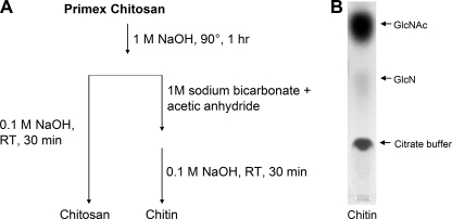
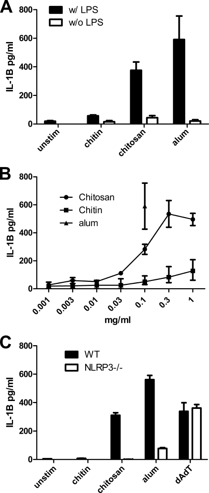
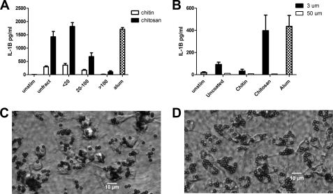
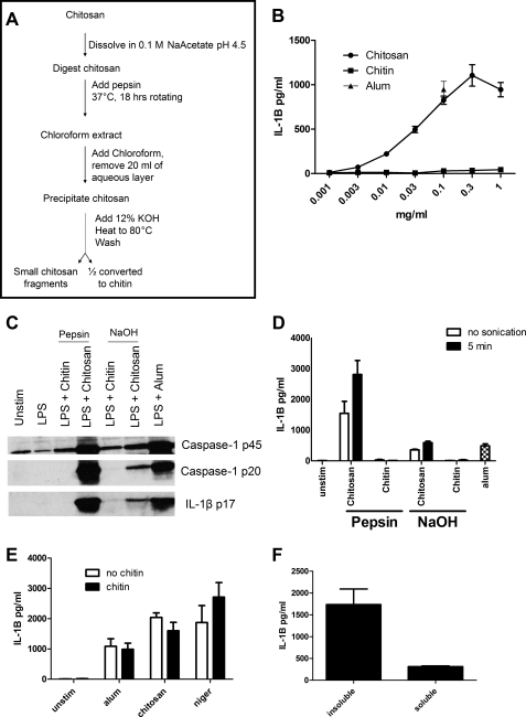
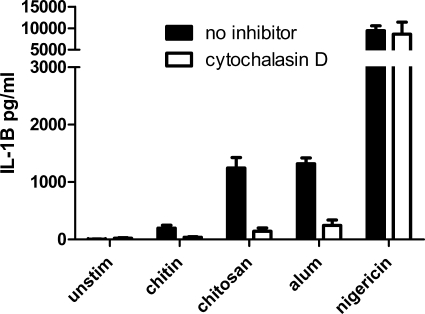
Chitin is an abundant polysaccharide found in fungal cell walls, crustacean shells, and insect exoskeletons. The immunological properties of both chitin and its deacetylated derivative chitosan are of relevance because of frequent natural exposure and their use in medical applications. Depending on the preparation studied and the end point measured, these compounds have been reported to induce allergic responses, inflammatory responses, or no response at all. We prepared highly purified chitosan and chitin and examined the capacity of these glycans to stimulate murine macrophages to release the inflammasome-associated cytokine IL-1β. We found that although chitosan was a potent NLRP3 inflammasome activator, acetylation of the chitosan to chitin resulted in a near total loss of activity. The size of the chitosan particles played an important role, with small particles eliciting the greatest activity. An inverse relationship between size and stimulatory activity was demonstrated using chitosan passed through size exclusion filters as well as with chitosan-coated beads of defined size. Partial digestion of chitosan with pepsin resulted in a larger fraction of small phagocytosable particles and more potent inflammasome activity. Inhibition of phagocytosis with cytochalasin D abolished the IL-1β stimulatory activity of chitosan, offering an explanation for why the largest particles were nearly devoid of activity. Thus, the deacetylated polysaccharide chitosan potently activates the NLRP3 inflammasome in a phagocytosis-dependent manner. In contrast, chitin is relatively inert.


Figures

FIGURE 1.
FIGURE 1.
Chitin and chitosan purification and TLC analysis. A, commercial chitosan was treated with 1 m sodium hydroxide. A portion of the chitosan was then converted to chitin by suspension in sodium bicarbonate with acetic anhydride to drive the acetylation reaction. Both preparations were then further purified in 0.1 m sodium hydroxide. B, to assess the efficacy of the acetylation reaction, the chitin was digested to monomers by T. viride chitinase and analyzed by TLC.
FIGURE 2.
FIGURE 2.
Inflammasome activation stimulated by chitin and chitosan. A, BMMΦ (1 × 10⁵/well) were primed for 3 h with 100 ng/ml LPS or left unprimed and then stimulated for 6 h with alum (0.1 mg/ml) or with the chitosan and chitin (0.1 mg/ml) preparations generated as in Fig. 1. Supernatants were assayed for the inflammasome cytokine IL-1β by ELISA. Data are means ± S.E. of four independent experiments, each performed in triplicate. p < 0.001 comparing primed chitin to primed chitosan, unprimed alum to primed alum, and unprimed chitosan to primed chitosan, as analyzed by two-way ANOVA. B, dose response curve of chitin and chitosan stimulating BMMΦ (1 × 10⁵/well) after they were primed for 3 h with 100 ng/ml LPS. Data are means ± S.E. of four independent experiments, each performed in triplicate. C, IL-1β production from stimulated WT and NLRP3−/− macrophages was compared. Chitin and chitosan were used at 1 mg/ml. Alum (1 mg/ml) and dAdT (2 μg/ml), which stimulate the NLRP3 and AIM2 inflammasomes, respectively, served as controls. IL-1β release was significantly reduced in NLRP3−/− macrophages stimulated with alum and chitosan. Data are means ± S.E. of a representative of two independent experiments, each performed in triplicate. p < 0.001 comparing WT macrophages and NALP3−/− macrophages stimulated by chitosan or alum as analyzed by two-way ANOVA.
FIGURE 3.
FIGURE 3.
The effect of particle size on inflammasome activation. A, chitosan and chitin preparations prepared as in Fig. 1 were sonicated and then size-fractionated through 100-μm and 20-μm filters. BMMΦ (1 × 10⁵/well) were primed with LPS and then stimulated with chitosan or chitin particles (1 mg/ml) that were left unfractionated (unfract) or size-fractionated as indicated. IL-1β was analyzed by ELISA. Data are means ± S.E. of three independent experiments, each performed in triplicate. p < 0.001 comparing unfractionated chitosan to 20–100 chitosan and >100 chitosan fractions, and between the <20 chitosan fraction and the 20–100 and >100 chitosan fractions, analyzed by two-way ANOVA. B, LPS-primed BMMΦ (1 × 10⁵/well) were left unstimulated (Unstim) or incubated for 6 h with the indicated size and type of beads (1 mg/ml). Alum (1 mg/ml) served as a positive control. Supernatants were analyzed for IL-1β by ELISA. Data are means ± S.E. of three independent experiments, each performed in triplicate. p < 0.01 comparing 3-μm chitosan beads and 50-μm chitosan beads by two-way ANOVA. Shown are representative photomicrographs of BMMΦ following 30-min incubation with 3-μm chitin-coated (C) and chitosan-coated (D) beads demonstrating robust phagocytosis of both types of glycan-coated beads.
FIGURE 4.
FIGURE 4.
Effect of pepsin digestion of chitosan on inflammasome activation. Following the procedure outlined in A, chitosan was digested with pepsin and then half was converted to chitin. B, dose curve of the pepsin-treated chitin and chitosan-stimulating BMMΦ (1 × 10⁵/well) after they were primed for 3 h with 100 ng/ml LPS. Data are means ± S.E. of four independent experiments, each performed in triplicate. p < 0.01 comparing chitin and chitosan at any concentration ≥ 0.1 mg/ml as analyzed by unpaired t test. C, BMMΦ (1.5 × 10⁶/well) were primed for 3 h with 100 ng/ml LPS and then stimulated with alum (0.1 mg/ml) or chitin and chitosan derived from the procedure outlined in Fig. 4A (pepsin) or the procedure outlined in Fig. 1A (NaOH). Supernatants were then collected and analyzed for caspase-1 and IL-1B by immunoblot. Caspase-1 p20 and IL-1B p17 represent the mature forms and indicate an active inflammasome, whereas caspase-1 p45 is an inactive proform of caspase-1. D, BMMΦ (1 × 10⁵/well) were primed as in B and then stimulated with alum or chitin and chitosan derived from the procedure outlined in Fig. 4A (pepsin) or the procedure outlined in Fig. 1A (NaOH). The chitin and chitosan preparations were left unsonicated (no sonication) or sonicated for 5 min (5 min). All stimuli were added at a concentration of 0.1 mg/ml. Supernatants were analyzed by ELISA for IL-1β. Data are means ± S.E. of three independent experiments, each performed in triplicate. p < 0.001 comparing no sonication and 5-min sonication of pepsin chitosan by two-way ANOVA. E, BMMΦ (1 × 10⁵/well) were primed as in B. Two hours later, wells either received 0.1 mg/ml chitin or were left without chitin treatment (no chitin). One hour later, cells were left unstimulated (unstim) or stimulated for 6 h with alum (0.1 mg/ml), or chitosan (0.1 mg/ml), or 1 h with nigericin (2.5 μm). Supernatants were analyzed by ELISA for IL-1β. Data are mean ± S.E. of two independent experiments, each performed in triplicate. F, BMMΦ (1 × 10⁵/well) were primed as in B. Insoluble suspended chitosan and chitosan that had been solubilized in acetic acid were diluted in media and added to cells. Supernatants were analyzed by ELISA for IL-1β. Data are means ± S.E. of two independent experiments, each performed in triplicate. p < 0.01 comparing insoluble with soluble chitosan by two-tailed unpaired t test.
FIGURE 5.
FIGURE 5.
Inhibition of phagocytosis blocks inflammasome activation. BMMΦ (1 × 10⁵/well) were primed for 3 h with 100 ng/ml LPS. The BMMΦ were treated with 1 μg/ml cytochalasin D to inhibit phagocytosis 10 min prior to addition of stimuli. After 1 h stimulation with nigericin (2.5 μm), or 6 h with alum (0.1 mg/ml), chitin (0.1 mg/ml), and chitosan (0.1 mg/ml), supernatants were collected and analyzed by ELISA. Data are means ± S.E. of three experiments performed in triplicate. p < 0.001 comparing cytokine concentrations with and without cytochalasin D following stimulation with alum and chitosan, analyzed by two-tailed unpaired t test.

References

    1. Boot R. G., Renkema G. H., Verhoek M., Strijland A., Bliek J., de Meulemeester T. M., Mannens M. M., Aerts J. M. (1998) J. Biol. Chem. 273, 25680–25685 - PubMed
    1. Neville A. C., Parry D. A., Woodhead-Galloway J. (1976) J. Cell Sci. 21, 73–82 - PubMed
    1. Boot R. G., Blommaart E. F., Swart E., Ghauharali-van der Vlugt K., Bijl N., Moe C., Place A., Aerts J. M. (2001) J. Biol. Chem. 276, 6770–6778 - PubMed
    1. Fuhrman J. A., Piessens W. F. (1985) Mol. Biochem. Parasitol. 17, 93–104 - PubMed
    1. Shahabuddin M., Kaslow D. C. (1994) Exp. Parasitol. 79, 85–88 - PubMed

Publication types

MeSH terms

LinkOut - more resources