. 2011 Oct 21;286(42):36852-63.
doi: 10.1074/jbc.M111.219931. Epub 2011 Aug 23.

InvA protein is a Nudix hydrolase required for infection by pathogenic Leptospira in cell lines and animals

Affiliations

InvA protein is a Nudix hydrolase required for infection by pathogenic Leptospira in cell lines and animals

Yihui Luo et al. J Biol Chem. .

Retraction in

Abstract

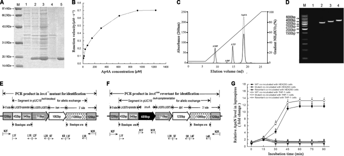
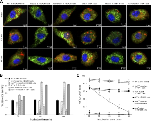
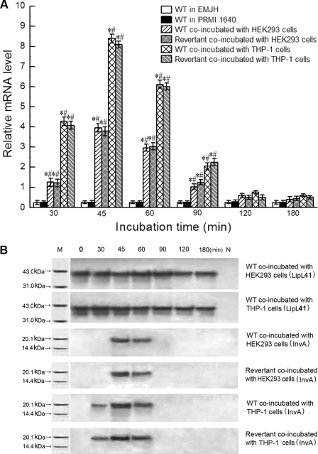
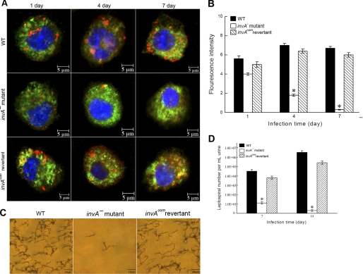
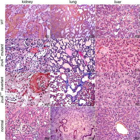
Leptospirosis caused by pathogenic species of the genus Leptospira is a re-emerging zoonotic disease, which affects a wide variety of host species and is transmitted by contaminated water. The genomes of several pathogenic Leptospira species contain a gene named invA, which contains a Nudix domain. However, the function of this gene has never been characterized. Here, we demonstrated that the invA gene was highly conserved in protein sequence and present in all tested pathogenic Leptospira species. The recombinant InvA protein of pathogenic L. interrogans strain Lai hydrolyzed several specific dinucleoside oligophosphate substrates, reflecting the enzymatic activity of Nudix in Leptospira species. Pathogenic leptospires did not express this protein in media but temporarily expressed it at early stages (within 60 min) of infection of macrophages and nephric epithelial cells. Comparing with the wild type, the invA-deficient mutant displayed much lower infectivity and a significantly reduced survival rate in macrophages and nephric epithelial cells. Moreover, the invA-deficient leptospires presented an attenuated virulence in hamsters, caused mild histopathological damage, and were transmitted in lower numbers in the urine, compared with the wild-type strain. The invA revertant, made by complementing the invA-deficient mutant with the invA gene, reacquired virulence similar to the wild type in vitro and in vivo. The LD(50) in hamsters was 1000-fold higher for the invA-deficient mutant than for the invA revertant and wild type. These results demonstrate that the InvA protein is a Nudix hydrolase, and the invA gene is essential for virulence in pathogenic Leptospira species.


Figures

FIGURE 1.
FIGURE 1.
Expression of leptospiral rInvA, identification of invA mutant and invAcom revertant, and Nudix hydrolase activity of the rInvA. A, expression and purification of rInvA protein of wild type of L. interrogans strain Lai. Lane M, protein marker (BioColor); lane 1, pET42a without invA gene insertion; lane 2, expressed rInvA induced with 1.0 mm IPTG at 28 °C; lanes 3 and 4, presence of rInvA in the bacterial supernatant and precipitate after ultrasonication; lane 5, rInvA purified by Ni-NTA affinity chromatography. B, Michaelis-Menten plot for the hydrolysis of Ap4A caused by the leptospiral rInvA. C, HPLC elution profiles of products of Ap4A caused by the leptospiral rInvA. A reaction mixture containing 50 mm Tris-HCl, pH 9.0, 1 mm substrate, 5 mm Mg2+ with or without 0.05 μm of the leptospiral rInvA was incubated for 20 min at 37 °C. The samples were analyzed on a 1-ml Resource Q column at a flow rate of 2 ml/min in 35 mm NH4HCO3, pH 9.6. The products were eluted and separated by an 18-min gradient elution from 5% to 100% with 0.7 m NH4HCO3. The proportion of hydrolyzed substrate was calculated by dividing the peak areas from the samples lacking the leptospiral rInvA (dotted line) by the samples containing the leptospiral rInvA (solid line). D, PCR results for identification of the invA mutant and invAcom revertant. Lane M, DNA ladder (TaKaRa); lane 1, blank control; lanes 2, the amplicon (3506 bp) of LA3978-invA-LA3976-LA3975–1086-bp segment (3266 bp) plus two extending regions from the 5′ (120 bp) and 3′ sides (120 bp) from wild type of L. interrogans strain Lai as the control; lanes 3, the amplicon (4080 bp) of LA3978-LA3976-LA3975-kan-1086bp segment (3840 bp) plus the two 120-bp extending regions from the invA mutant for identification; lanes 4, the amplicon (4741 bp) of LA3978-invA-LA3976-LA3975-spc-1086bp segment (4501 bp) plus the two 120-bp extending regions from the invAcom revertant for identification. The amplicons in lanes 2–4 were amplified using the same primers N2-F/N2-R. E, schematic diagram of the sequencing result of the invA mutant. The positions of PCR primers were marked below. F, schematic diagram of the sequencing result of the invAcom revertant. The positions of PCR primers are marked below. G, relative ApnN levels in the invA mutant, invAcom revertant, and wild type (WT) of L. interrogans strain Lai during infection. The relative ApnN levels in the invA mutant, invAcom revertant, and wild type L. interrogans strain Lai during infection of host cells for the indicated incubation times are expressed as -fold changes compared with each of the leptospires in media before infection (incubation time = 0), which was set as 1.0. * and #: p < 0.05 versus the wild type.
FIGURE 2.
FIGURE 2.
Roles of the leptospiral InvA in invasion and survival in host cells. A, typical microphotographs of HEK293 and THP-1 cells co-incubated with the invA mutant, invAcom revertant, and wild type of L. interrogans strain Lai for the indicated times. Blue: nuclei; green: cytoplasm; red: leptospires. B, fluorescence intensity reflects internalization levels of the invA mutant, invAcom revertant, and wild type of L. interrogans strain Lai in infected HEK293 and THP-1 cells for the indicated times. Statistical data from experiments such as shown in A are shown. 100 cells were analyzed to quantify for each the values of fluorescence signal intensity. * and #: p < 0.05 versus both the wild type and invAcom revertant. C, colony-forming units of the intracellular leptospires recovered from THP-1 and HEK293 cells infected for the indicated times. * and #: p < 0.05 versus both the wild type and invAcom revertant.
FIGURE 3.
FIGURE 3.
Increase of invA transcription and expression levels of invAcom revertant and wild type (WT) of L. interrogans strain Lai during infection. A, mRNA levels of the invAcom revertant and wild type during infection of HEK293 and THP-1 cells for the indicated times. mRNA levels of wild-type L. interrogans strain Lai in EMJH and PRMI 1640 media served as controls. * and #: p < 0.05 versus leptospires in both EMJH and RPMI 1640 media, respectively. B, InvA protein expression levels of the invAcom revertant and wild type during infection of HEK293 and THP-1 cells for the indicated times. Membrane lipoprotein LipL41 expression levels of wild-type L. interrogans strain Lai served as controls. M, protein marker (BioColor); N, blank controls.
FIGURE 4.
FIGURE 4.
Survival of the invA mutant, invAcom revertant, and wild-type (WT) of L. interrogans strain Lai in hamsters. A, leptospires of the invA mutant, invAcom revertant, and wild type in hamster monocytes for the indicated times. Blue: nucleus; green: cytoplasm; red: leptospires. B, statistical summary of fluorescence intensity (as in A) reflecting internalization levels of the invA mutant, invAcom revertant, and wild type in hamster monocytes for the indicated times. 100 cells of each strain were analyzed to quantify the fluorescence signal intensity. *: p < 0.05 versus the wild type and invAcom revertant. C, leptospires in urine of hamsters infected with the invA mutant, invAcom revertant, and wild-type (Fontana silver staining). D, counts of leptospires of the invA mutant, invAcom revertant, and wild type in urine of hamsters at the indicated times. *: p < 0.05 versus the wild type and invAcom revertant.
FIGURE 5.
FIGURE 5.
Histopathological damage in hamsters infected with the invA mutant, invAcom revertant, and wild-type (WT) of L. interrogans strain Lai. Serious congestion and multiple focal necrosis of nephric tubular epithelia in kidney, evident hemorrhaging and inflammatory cell infiltration in lung, and extensive hepatocyte necrosis in liver occurred in hamsters infected with 106 leptospires per animal of the invAcom revertant or the wild type. Conversely, mild cellular edema in kidney, inflammatory cell infiltration in lung, and slight granular degeneration in liver occurred in hamsters infected with the same number of the invA mutant.

Similar articles

Cited by

References

    1. Bharti A. R., Nally J. E., Ricaldi J. N., Matthias M. A., Diaz M. M., Lovett M. A., Levett P. N., Gilman R. H., Willig M. R., Gotuzzo E., Vinetz J. M. (2003) Lancet Infect. Dis. 3, 757–771 - PubMed
    1. Vijayachari P., Sugunan A. P., Shriram A. N. (2008) J. Biosci. 33, 557–569 - PubMed
    1. Djadid N. D., Ganji Z. F., Gouya M. M., Rezvani M., Zakeri S. (2009) Diagn. Microbiol. Infect. Dis. 63, 251–256 - PubMed
    1. Chidambaram N., Ramanathan M., Anandi V., Sasikala S., Innocent D. J., Sarayu L. (2007) J. Commun. Dis. 39, 105–108 - PubMed
    1. Lee S. H., Kim S., Park S. C., Kim M. J. (2002) Infect. Immun. 70, 315–322 - PMC - PubMed

Publication types

MeSH terms