. 2011 Oct 21;286(42):36724-38.
doi: 10.1074/jbc.M111.277673. Epub 2011 Aug 23.

Auto-activation of c-JUN gene by amino acid deprivation of hepatocellular carcinoma cells reveals a novel c-JUN-mediated signaling pathway

Affiliations

Auto-activation of c-JUN gene by amino acid deprivation of hepatocellular carcinoma cells reveals a novel c-JUN-mediated signaling pathway

Lingchen Fu et al. J Biol Chem. .

Abstract

Mammalian cells respond to protein or amino acid (AA) limitation by activating a number of signaling pathways, collectively referred to as the AA response (AAR), that modulate a range of cellular functions, including transcriptional induction of target genes. This study demonstrates that in hepatocellular carcinoma cells, expression of c-JUN, JUN-B, c-FOS, and FOS-B was induced by the AAR, whereas JUN-D, FRA-1, and FRA-2 were not. Of the four activated FOS/JUN members, c-JUN made the largest contribution to the induction of several known AAR target genes. For several human liver, prostate, and ovarian cell lines, the AAR-induced increase in c-JUN expression was greater in transformed cells compared with nontransformed counterparts, an effect independent of cell growth rate. Thus far, the best characterized AA-responsive genes are all transcriptionally activated by ATF4, but the AAR-dependent induction of c-JUN transcription was ATF4-independent. The increased expression of c-JUN was dependent on ATF2 and on activation of the MEK-ERK and JNK arms of the MAPK signaling pathways. Formation of c-JUN-ATF2-activated heterodimers was increased after AA limitation, and c-JUN or ATF2 knockdown suppressed the induction of c-JUN and other AAR target genes. AA deprivation triggers a feed-forward process that involves phosphorylation of existing c-JUN protein by JNK and subsequent auto-activation of the c-JUN gene by recruitment of c-JUN and ATF2 to two AP-1 sites within the proximal promoter. The results document the novel observation that AP-1 sequences within the c-JUN gene can function as transcriptional amino acid-response elements.


Figures
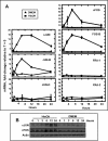
FIGURE 1.
FIGURE 1.
Expression of JUN/FOS family members in HepG2 hepatocellular carcinoma cells after activation of AAR. A, mRNA content of individual JUN and FOS family members were analyzed by qPCR in HepG2 human hepatoma cells incubated in control medium (DMEM) or medium containing 2 mm HisOH for 0–24 h. GAPDH mRNA, which is unchanged by the AAR, was used as the internal control. The results are the averages ± S.D. of three or more assays. Where not shown, the standard deviation bars are contained within the symbol. B, after incubating the cells as described above, a 40-μg aliquot of whole cell extract was subjected to gel electrophoresis, and then the blots were probed with primary antibody against c-JUN, c-FOS, or actin.
FIGURE 2.
FIGURE 2.
Induction of c-JUN expression by the AAR occurs by an ATF4-independent mechanism. A, diagram is shown of the human c-JUN genomic structure and the sites (P1, nt −9949/−9895; P2, nt −484/−400; and P3, nt +12446/+12587) at which primers were used to analyze protein binding by ChIP. The transcription start site for the c-JUN mRNA is indicated with the arrow, and the protein coding sequence of this intronless gene is labeled as CDS, and a possible CARE site is indicated. HepG2 cells were incubated in control (DMEM) medium or medium containing 2 mm HisOH for 8 h. To test for enrichment of ATF4 protein binding at the indicated region of the gene, ChIP assays were performed, and the data were plotted as the ratio to the value obtained with a 1:20 dilution of input DNA. As a positive control, ATF4 binding to the AARE in the ASNS promoter was monitored. Each condition was analyzed in triplicate and repeated in at least two independent experiments for which, an asterisk indicates p ≤ 0.05. B, HepG2 cells, transfected with control siRNA (si-Con) or siRNA against ATF4 (si-ATF4), were incubated in DMEM ± 2 mm HisOH for 8 h, and then c-JUN or ASNS mRNA content was analyzed by qPCR. ATF4 protein content was also analyzed to monitor the knockdown efficiency. An asterisk indicates a p value of <0.05 when comparing the si-ATF4 + HisOH values to the control siRNA + HisOH values. C, HEK293-ATF4 cells, which stably express a tetracycline-inducible ATF4, were treated with the indicated amount of tetracycline for 8 h, and the c-JUN and ATF4 protein content was analyzed by immunoblot with actin serving as a loading control.
FIGURE 3.
FIGURE 3.
JNK and MEK-ERK signaling contributes to c-JUN induction by AAR. A, for AA deprivation in the presence of a JNK or MEK inhibitor, HepG2 cells were pretreated for 1 h with the indicated concentration and then incubated with the inhibitor in DMEM (D) or DMEM ± 2 mm HisOH (H) for an additional 4 h. Whole cell extracts were analyzed for the indicated proteins by immunoblotting. B, HEK293 cells were transiently transfected with an expression plasmid encoding constitutively active MEK or, as a negative control, green fluorescent protein (Ctrl) and then 36 h later they were incubated in fresh DMEM ± HisOH for 8 h. The total c-JUN protein content was analyzed by immunoblot, and to document the effectiveness of the MEK expression, p-ERK protein content was measured as well, with actin serving as a loading control. The c-JUN mRNA content was analyzed by qPCR, and an asterisk indicates that the value for MEK-induced expression is statistically greater than the corresponding control (p ≤ 0.05). C, HepG2 cells were transfected with GFP (Ctrl) or a constitutively active MEK (MEK), and 36 h later, one-half of the cells were pretreated with MEK inhibitor for 1 h. The cells were then transferred to either DMEM (D) or DMEM + 2 mm HisOH (H) with or without the inhibitor for an additional 4 h. Whole cell extracts were probed by immunoblotting with antibodies specific for the indicated proteins.
FIGURE 4.
FIGURE 4.
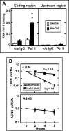
c-JUN mRNA is increased by transcription and stabilization following AAR activation. A, HepG2 cells were maintained in DMEM ± 2 mm HisOH for 6 h, and then RNA pol II binding to the c-JUN gene was assayed by ChIP and qPCR to estimate transcription activity, as described under “Experimental Procedures.” Nonspecific IgG and primers for a 5′ upstream region of the gene (nt −9949) were used as negative controls. The asterisk indicates a p ≤ 0.05 when comparing the DMEM + HisOH values to those for DMEM alone. B, to measure mRNA half-life, HepG2 cells were incubated in DMEM ± 2 mm HisOH for 4 h and then transferred to control medium (DMEM) or DMEM + 2 mm HisOH, both containing 5 μm actinomycin D (ActD). Total RNA was isolated from triplicate samples at the times indicated, and qPCR was performed to quantify the c-JUN and ASNS mRNA. The data were plotted as the logarithm of mRNA content versus time after transfer to the ActD-containing media. Where not shown, the standard deviation bars are contained within the symbol.
FIGURE 5.
FIGURE 5.
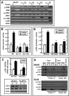
c-JUN expression is regulated by c-JUN and ATF2 through ERK and JNK signaling. A, HepG2 cells were incubated for 0–6 h in DMEM ± 2 mm HisOH, and the whole cell extracts were subjected to immunoblotting with the indicated antibodies. B, HepG2 cells were transiently transfected with a Firefly luciferase reporter construct driven by a fragment of the c-JUN gene (nt −370 to +731). As indicated, constructs in which either of the AP-1 sites (nt −190 or −71) was deleted were also tested. After transfection, the cells were incubated in DMEM ± 2 mm HisOH for 3 h prior to analysis of luciferase activity as a measure of c-JUN promoter function. C, HepG2 cells were transfected with expression plasmids encoding a sh-Control (sh-Con) or a sequence specific against ATF2 (sh-ATF2). After culture for 48 h, transfected cells were then incubated in DMEM ± HisOH for 4 h. The c-JUN mRNA content was measured by qPCR (C, upper panel), and the protein content of c-JUN and ATF2 was analyzed by immunoblotting using actin as a loading control (C, lower panel). D, HepG2 cells were co-transfected with a c-JUN promoter/luciferase reporter plasmid and expression plasmids encoding an sh-Control (sh-Con) sequence or an sh-RNA sequence specific for either c-JUN (sh-c-JUN) or ATF2 (sh-ATF2). After culture for 48 h, transfected cells were incubated in DMEM ± HisOH for 3 h. Cell extracts were assayed and normalized for luciferase activity as described under “Experimental Procedures.” The data are presented as the averages ± S.D. for at least three independent assays, and each experiment was repeated at least twice. An asterisk indicates a p value of ≤0.05 relative to the sh-Con (+ HisOH). E, DAPA analysis of both AP-1 sequences within the c-JUN promoter was performed as described under “Experimental Procedures.” Nuclear protein extracts from HepG2 cells maintained for 4 h in DMEM (D) or DMEM + 2 mm HisOH (H) were incubated with Sepharose-bound AP-1 DNA sequence alone (Probe71 or Probe 190) or with a five times amount of the same sequence as an unbound DNA competitor “Probe + Comp”). The bound proteins were subjected to immunoblotting along with an aliquot of the starting nuclear extract (Input) and an incubation that did not contain any protein extract (No extract). Equal loading of the samples was established by Fast Green staining of the blot (not shown).
FIGURE 6.
FIGURE 6.
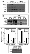
ATF2 heterodimerizes with c-JUN and contributes to the induction of AAR-dependent genes. A, protein content of total ATF2 and c-JUN, as well as their phosphorylated forms, was analyzed by immunoblot in extracts from HepG2 human hepatoma cells incubated in DMEM (D) ± 2 mm HisOH (H) for 0–24 h. (The actin blot and the total c-JUN blot are the same as those in Fig. 1B.) B, HepG2 cells were incubated in DMEM ± 2 mm HisOH for 8 h and then analyzed for protein-protein interactions by immunoprecipitation (IP-Ab) and immunoblotting (IB-Ab) with the indicated antibodies, as described under “Experimental Procedures.” C, HepG2 cells were co-transfected with a CHOP- or an ASNS-driven Firefly luciferase reporter plasmid and expression plasmids encoding an sh-Control (sh-Con) or an sh-ATF2 sequence. After culture for 48 h and incubation in DMEM ± HisOH for 15 h, the promoter activity was monitored by assaying luciferase activity that was normalized to cell protein content. The luciferase data are presented as the averages ± S.D. for at least three independent assays, and each experiment was repeated at least twice. An asterisk indicates a p value of ≤0.05 when comparing the sh-ATF2 + HisOH value to the sh-Control + HisOH value. To indicate the level of ATF2 knockdown, the protein content of total ATF2 protein was analyzed by immunoblot using actin as a loading control.
FIGURE 7.
FIGURE 7.
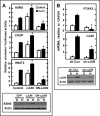
c-JUN impacts the regulated expression of downstream AAR target genes. A, HepG2 cells were co-transfected with the indicated genomic promoter fragment/Firefly luciferase reporter plasmid (ASNS, CHOP, or SNAT2) and expression plasmids for wild type c-JUN or a dominant negative c-JUN (DN-c-JUN). After a 24-h incubation, transfected cells were treated with 2 mm HisOH (H) for 15 h, and cell extracts were assayed for Firefly luciferase activity, and the results normalized to cell protein. As a positive control for c-JUN function, the endogenous protein content of ASNS was measured by immunoblotting. D, DMEM. B, HepG2 cells were transfected with a plasmid encoding a control sh-RNA (sh-Con) or an sh-RNA against c-JUN (sh-c-JUN). After a 40-h selection in puromycin to enrich for transfected cells, they were incubated with DMEM ± 2 mm HisOH for 4 h. The mRNA content of c-JUN and FOXA2 was analyzed by qPCR, and the protein content of c-JUN was analyzed by immunoblot using actin as a loading control. The data are presented as the averages ± S.D. for at least three independent assays, and each experiment was repeated at least twice. The statistical significance from the control is indicated with an asterisk (p ≤ 0.05) when comparing the experimental treatment to the appropriate control (DMEM versus DMEM + c-JUN or DN-c-JUN; HisOH alone versus HisOH + c-JUN or DN-c-JUN).
FIGURE 8.
FIGURE 8.
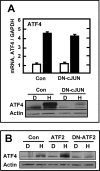
AAR-induced c-JUN enhances ATF4 protein levels. A, HepG2 cells were transfected with empty plasmid (Con) or a plasmid encoding DN-c-JUN. After a 48-h incubation, the transfected cells were incubated in DMEM (D) ± 2 mm HisOH (H) for 4 h and then either ATF4 mRNA or ATF4 and c-JUN protein content was analyzed. B, HepG2 cells were transfected with plasmids encoding wild type ATF2 or DN-ATF2. After a 40-h selection in puromycin to enrich the transfected cells, they were incubated in DMEM ± 2 mm HisOH for 4 h, and whole cell extracts were analyzed for ATF4 and actin protein by immunoblotting.
FIGURE 9.
FIGURE 9.
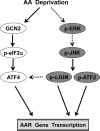
Working model for the ATF4-dependent and c-JUN-dependent AA signaling pathways. The GCN2-eIF2α-ATF4 pathway that culminates in ATF4 binding to CARE sites has been well documented (7). The novel information in this study illustrates the existence of a ERK-JNK-c-JUN/ATF2 pathway that functions independently of ATF4 by assembling c-JUN-c-JUN or c-JUN-ATF2 dimers at AP-1 sites. Although not indicated for clarity, it is known that ATF2 also participates in activating ATF4-dependent genes by catalyzing histone acetylation (16, 48). The dashed lines indicate that the mechanism is unknown.

Similar articles

Cited by

References

    1. Lillycrop K. A., Phillips E. S., Jackson A. A., Hanson M. A., Burdge G. C. (2005) J. Nutr. 135, 1382–1386 - PubMed
    1. Morgane P. J., Austin-LaFrance R., Bronzino J., Tonkiss J., Díaz-Cintra S., Cintra L., Kemper T., Galler J. R. (1993) Neurosci. Biobehav. Rev. 17, 91–128 - PubMed
    1. Morley J. E. (2009) Curr. Opin. Clin. Nutr. Metab. Care 12, 607–610 - PubMed
    1. Pedrini M. T., Levey A. S., Lau J., Chalmers T. C., Wang P. H. (1996) Ann. Intern. Med. 124, 627–632 - PubMed
    1. Zimmerman J. A., Malloy V., Krajcik R., Orentreich N. (2003) Exp. Gerontol. 38, 47–52 - PubMed

Publication types

MeSH terms