. 2011 Oct 7;286(40):34941-50.
doi: 10.1074/jbc.M111.286427. Epub 2011 Aug 23.

Molecular mechanisms for synchronized transcription of three complement C1q subunit genes in dendritic cells and macrophages

Affiliations

Molecular mechanisms for synchronized transcription of three complement C1q subunit genes in dendritic cells and macrophages

Guobao Chen et al. J Biol Chem. .

Abstract

Hereditary homozygous C1q deficiency is rare, but it almost certainly causes systemic lupus erythematosus. On the other hand, C1q levels can decline in systemic lupus erythematosus patients without apparent C1q gene defects and the versatility in C1q production is a likely cause. As an 18-subunit protein, C1q is assembled in a 1:1:1 ratio from three different subunits. The three human C1q genes are closely bundled on chromosome 1 (C1qA-C1qC-C1qB) and their basal and IFNγ-stimulated expression, largely restricted to macrophages and dendritic cells, is apparently synchronized. We cloned the three gene promoters and observed that although the C1qB promoter exhibited basal and IFNγ-stimulated activities consistent with the endogenous C1qB gene, the activities of the cloned C1qA and C1qC promoters were suppressed by IFNγ. To certain extents, these were corrected when the C1qB promoter was cloned at the 3' end across the luciferase reporter gene. A 53-bp element is essential to the activities of the C1qB promoter and the transcription factors PU.1 and IRF8 bound to this region. By chromatin immunoprecipitation, the C1qB promoter was co-precipitated with PU.1 and IRF8. shRNA knockdown of PU.1 and IRF8 diminished C1qB promoter response to IFNγ. STAT1 instead regulated C1qB promoter through IRF8 induction. Collectively, our results reveal a novel transcriptional mechanism by which the expression of the three C1q genes is synchronized.


Figures

FIGURE 1.
FIGURE 1.
Basal and IFNγ-induced expression of C1qA, C1qB, and C1qC mRNA in cultured DCs (left) and macrophages (right). DCs and macrophages were cultured from blood monocytes and were, at day 6, cultured for a further 24 h either untreated (PBS) or treated with human IFNγ (100 ng/ml). RNA was isolated for real-time PCR using primers specific for C1qA, C1qB, and C1qC. Results were normalized to the levels of β-actin mRNA in each experiment, and data were presented as folds of C1q mRNA induction by IFNγ taking that detected in untreated cells as 1. Conventional PCR was also performed, and the PCR products were detected on 1% agarose gels.
FIGURE 2.
FIGURE 2.
Basal and IFNγ-stimulated activities of the C1qA, C1qB, and C1qC gene promoters. The anticipated promoter regions for C1qA, C1qB, and C1qC were specified and cloned into the pGL3-basic vector (A). The cloned C1qA (B), C1qB (C), and C1qC (D) gene promoters were transfected into RAW264.7 cells to measure the basal activities, and cells were treated with mouse IFNγ (10 ng/ml) to measure IFNγ-stimulated activities. After 5′ deletions, the shortened promoters were examined similarly. In each experiment, a Renilla luciferase reporter plasmid under the β-actin promoter was co-transfected to normalize the firefly luciferase activities derived from C1q promoters. Data were presented as means ± S.D. of triplicate experiments.
FIGURE 3.
FIGURE 3.
Predicted cis-acting elements in the B273 promoter. The sequence of the −273-bp C1qB promoter (B273) was analyzed using MatInspector software, and the predicted cis-acting elements were highlighted. Two noticeable elements identified are the GAS-ISRE chimeric site proximal to the TSS (+1) and an upstream c-Rel site.
FIGURE 4.
FIGURE 4.
Identification of a 53-bp IFNγ-stimulated cis-acting element in the C1qB promoter. A, further 5′ deletions of the B273 promoter at −144, −133, −125, and −119 bp were made to identify the 5′ boundary of the IFNγ-stimulated region in the C1qB promoter. B, 3′ deletions were made at −28, −45, −72, −81, and −90 bp to identify the 3′ boundary of the IFNγ-stimulated site. C, illustration of site-directed mutagenesis of the B273 promoter to determine the involvement of specific nucleotides for B273 response to IFNγ. D, basal and IFNγ-stimulated activities of site-directed B273 mutants. Data were presented at mean ± S.D. of triplicate experiments.
FIGURE 5.
FIGURE 5.
Superseding regulation of 5′ C1qA and C1qC promoters by a 3′ B273 promoter. A, schematic of expression constructs in which the B273 promoter was cloned at the 3′ end of the luciferase reporter gene that either lacked a 5′ promoter (pGL3-B273) or were flanked by a 5′ C1qA (C1qA-B273), C1qC (C1qC-B273), or shortened C1qC (C258-B273) promoters. Control plasmids were also illustrated. B, the basal and IFNγ-stimulated activities of these constructs were compared with the control constructs. Data were presented as mean ± S.D. of triplicate experiments.
FIGURE 6.
FIGURE 6.
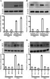
Roles of PU.1, IRF8, STAT1, and IRF1 in IFNγ stimulation of the C1qB promoter. RAW264.7 cells were transfected with the B273 promoter and also co-transfected with plasmids encoding shRNA for mouse IRF1 (A), STAT1 (B), IRF8 (IRF8–1, IRF8–2; C), PU.1 (PU.1-1, PU.1-2; D) or, as controls, with plasmids for scramble shRNA. Basal and IFNγ-stimulated activities were determined after the knockdown of these transcription factors. The knockdown of IRF1, STAT1, IRF8, and PU.1 expression was verified by Western blotting (upper panels). As a control, the expression of β-actin was monitored (lower panels). The luciferase data were presented as mean ± S.D. of triplicate experiments.
FIGURE 7.
FIGURE 7.
Binding of PU.1 and IRF8 to a 53-bp fragment of the C1qB promoter (−133 to −81 bp). This 53-bp DNA fragment was synthesized with 3′-biotin tag, which was then immobilized on streptavidin-Sepharose resins. Nuclear extract was prepared from untreated (PBS) and IFNγ-stimulated (IFNγ) RAW264.7 cells and then incubated with the immobilized 53-bp DNA fragment. After washing, the precipitated proteins were analyzed by Western blotting to detect IRF1, STAT1, IRF8, and PU.1 with specific antibodies. As a positive control, nuclear extract (10% of the input amount) was also included in the blots. Proteins precipitated with streptavidin-Sepharose without the 53-bp DNA fragment were used as negative controls.
FIGURE 8.
FIGURE 8.
STAT1 is required for IFNγ-stimulated IRF8 expression. IRF8 mRNA was detected in RAW264.7 cells after transfection with IRF8 shRNA (shIRF8–1 and shIRF8–2) or STAT1 shRNA plasmids (shSTAT1). As a control, the cells were transfected with scramble shRNA plasmid. Cells were either untreated (PBS) or IFNγ-stimulated (IFNγ) for 24 h and IRF8 mRNA was measured by real-time PCR. Data were presented as mean ± S.D. of triplicate experiments.
FIGURE 9.
FIGURE 9.
Endogenous PU.1 and IRF8 association with the C1q gene promoters. Macrophages were cultured from human blood monocytes and fixed with formaldehyde. Nuclear fraction was harvested and sonicated to generate nuclear lysate. The chromatin fragments were incubated with protein G-Sepharose immobilized with antibodies specific for IRF1, IRF8, STAT1, and PU.1. As negative controls, immunoprecipitation was performed with anti-His antibody or without antibody. Precipitated chromatin fragments were eluted, and DNA was extracted for PCR detection of specific C1qA, C1qB, and C1qC promoter regions. DNA isolated from sonicated nuclei before immunoprecipitation was used as a positive control. PCR products were examined on 1% agarose gels.
FIGURE 10.
FIGURE 10.
A proposed core promoter function for the C1qB promoter. The B273 promoter falls in the classic promoter region of its downstream C1qB gene. However, its upstream C1qA and C1qC genes lack the activities expected from results obtained with the endogenous genes. The fact that the B273 promoter is able to supersede these expected C1qA and C1qC gene promoters from the 3′ ends, across the luciferase reporter gene, suggests B273 regulation of C1qA and C1qC gene expression from the 3′ ends of these genes. This model explains the highly conserved chromosomal clustering and synchronized transcription of the three C1q genes.

References

    1. Reid K. B. (1986) Essays Biochem. 22, 27–68 - PubMed
    1. Walport M. J. (2001) N. Engl. J. Med. 344, 1058–1066 - PubMed
    1. Fujita T., Matsushita M., Endo Y. (2004) Immunol. Rev. 198, 185–202 - PubMed
    1. Colten H. R., Strunk R. C., Perlmutter D. H., Cole F. S. (1986) Ciba Found. Symp. 118, 141–154 - PubMed
    1. Müller W., Hanauske-Abel H., Loos M. (1978) J. Immunol. 121, 1578–1584 - PubMed

Publication types