. 2011 Oct 14;286(41):35456-35465.
doi: 10.1074/jbc.M111.236794. Epub 2011 Aug 23.

Histone 3 lysine 9 (H3K9) methyltransferase recruitment to the interleukin-2 (IL-2) promoter is a mechanism of suppression of IL-2 transcription by the transforming growth factor-β-Smad pathway

Affiliations

Histone 3 lysine 9 (H3K9) methyltransferase recruitment to the interleukin-2 (IL-2) promoter is a mechanism of suppression of IL-2 transcription by the transforming growth factor-β-Smad pathway

Yu Wakabayashi et al. J Biol Chem. .

Abstract

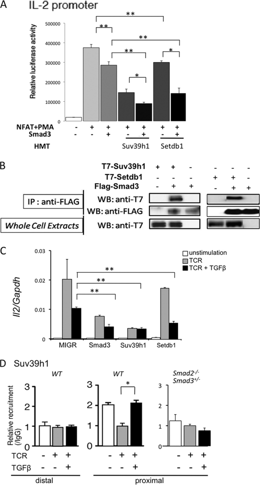
Suppression of IL-2 βproduction from T cells is an important process for the immune regulation by TGF-β. However, the mechanism by which this suppression occurs remains to be established. Here, we demonstrate that Smad2 and Smad3, two major TGF-β-downstream transcription factors, are redundantly essential for TGF-β-mediated suppression of IL-2 production in CD4(+) T cells using Smad2- and Smad3-deficient T cells. Both Smad2 and Smad3 were recruited into the proximal region of the IL-2 promoter in response to TGF-β. We then investigated the histone methylation status of the IL-2 promoter. Although both histone H3 lysine 9 (H3K9) and H3K27 trimethylation have been implicated in gene silencing, only H3K9 trimethylation was increased in the proximal region of the IL-2 promoter in a Smad2/3-dependent manner, whereas H3K27 trimethylation was not. The H3K9 methyltransferases Setdb1 and Suv39h1 bound to Smad3 and suppressed IL-2 promoter activity in collaboration with Smad3. Overexpression of Suv39h1 in 68-41 T cells strongly inhibited IL-2 production in response to T cell receptor stimulation irrespective of the presence or absence of TGF-β, whereas Setdb1 overexpression only slightly suppressed IL-2 production. Silencing of Suv39h1 by shRNA reverted the suppressive effect of TGF-β on IL-2 production. Furthermore, TGF-β induced Suv39h1 recruitment to the proximal region of the IL-2 promoter in wild type primary T cells; however, this was not observed in Smad2(-/-)Smad3(+/-) T cells. Thus, we propose that Smads recruit H3K9 methyltransferases Suv39h1 to the IL-2 promoter, thereby inducing suppressive histone methylation and inhibiting T cell receptor-mediated IL-2 transcription.


Figures

FIGURE 1.
FIGURE 1.
Smad2 and Smad3 are redundantly essential for TGF-β-mediated IL-2 suppression. A, naïve CD4+ cells from WT mice were stimulated with anti-CD3 and anti-CD28 antibodies in the presence or absence of TGF-β for indicated periods mRNA levels of IL-2 and Foxp3 were measured by real time RT-PCR. B, naïve CD4+ cells from LckCreSmad2fl/fl (Smad2−/−), Smad3−/−, or LckCre-Smad2fl/flSmad3+/− (Smad2−/−Smad3+/−) mice were stimulated with anti-CD3 and anti-CD28 in the presence (+) or absence (−) of TGF-β for 24 h. mRNAs were isolated from T cells, and microarray analysis was performed. Normalized Foxp3 and IL-2 mRNA levels in TGF-β (+) compared with TGF-β (−) are shown. C, FACS-sorted naïve CD4+ T cells stimulated with anti-CD3 and anti-CD28 with or without TGF-β1 at various concentrations. On day 3, cells were restimulated for 5 h with PMA and ionomycin. Brefeldin A was added to cultures <1 h after restimulation began. Cells were assessed for IL-2 production by intracellular staining. Data shown are representative of three independent experiments in triplicate with similar results.
FIGURE 2.
FIGURE 2.
Smad3 is recruited to the IL-2 promoter proximal regions where it suppresses IL-2 promoter activity. A, naïve T cells (7 × 10⁶) were stimulated with anti-CD3 and anti-CD28 antibodies (TCR) in the presence or absence of TGF-β for 24 h, and then a ChIP assay was performed using anti-Smad2 and Smad3 specific antibodies. The distal (−8.3 to −8.1) and proximal regions (−0.4 to −0.3) of the IL-2 promoter DNA were amplified and quantified by real-time PCR. Values are means + S.D. (n = 3) *, p < 0.05. B, IL-2 promoter activation by NF-AT and suppression by Smad3. The indicated length of the 5′-non-coding region of the IL-2 promoter was fused to luciferase and transfected into 293 cells together with NF-AT and Smad3 cDNA (1 μg). Transfected cells were stimulated with PMA, and luciferase activity was measured. **, p < 0.01. C, nucleic acid sequence and putative transcription factor binding sites. CAGA motifs are indicated. D, effect of mutations in CAGA motifs in −194 promoter on Smad3-mediated promoter suppression. 293 cells were transfected with the indicated reporters, NF-AT, and Smad3, and luciferase activity was then measured. **, p < 0.01.
FIGURE 3.
FIGURE 3.
DNA binding of Smads was not necessary for IL-2 promoter suppression. A, −194 IL-2 promoter was transfected into 293 cells together with NF-AT and Smad2 or Smad3 cDNA plasmids. After stimulation with PMA, luciferase activity was measured. Values are means + S.D. (n = 3). **, p < 0.01. B, effect of mutations in the DNA binding region of Smad3 on IL-2 promoter suppression. Left, WT or R74A mutant Smad3 cDNA and SBE-luciferase were transfected into 293 cells. After 48 h, luciferase activity was measured. Right, −194 IL-2 promoter-reporter was co-transfected with each mutant together with NF-AT. Expression levels of Smad3 proteins were determined by Western blotting with anti-FLAG antibody (lower panels). **, p < 0.01.
FIGURE 4.
FIGURE 4.
Histone modification in the IL-2 promoter region in response to TCR and TGF-β stimulation in primary T cells. A, trimethylation of H3K9, H3K27, and H3K4 in stimulated primary T cells from WT and Smad2- or Smad3-deficient mice as assessed through a ChIP assay. Primary CD4+ T cells from indicated mice were stimulated with anti-CD3 and anti-CD28 antibodies (TCR) in the presence or absence of TGF-β for 24 h. Histone trimethylation (H3K9me3, H3K27me3, and H3K4me2) on the proximal region of the IL-2 promoter (−0.3 to −0.4 kb upstream of the first ATG codon) was detected with ChIP assay using the indicated antibodies. ND, not determined. Values are means + S.D. (n = 3). *, p < 0.05; **, p < 0.01. B, K9 trimethylation of H3 (H3K9me3) at the IL-2 promoter was analyzed through a ChIP assay. Primary T cells were stimulated with anti-CD3 antibody in the presence or absence of TGF-β for 24 h. The upper panel shows the region amplified after immunoprecipitation with anti-H3K9me3 antibody. **, p < 0.01.
FIGURE 5.
FIGURE 5.
Histone methyltransferases interact with Smad3 and enhance IL-2 promoter suppression activity. A, 293 cells were transfected with −194 IL-2 promoter-reporter, NF-AT, and a small amount of Smad3 (0.1 μg) together with the indicated HMT cDNAs (1 μg). After stimulation with PMA, luciferase activity was measured. Values are means + S.D. (n = 3). *, p < 0.05; **, p < 0.01. B, binding of Smad3 to HMTs. 293 cells were transfected with the indicated plasmids and then lysed. After immunoprecipitation with anti-FLAG antibody, co-precipitated proteins were analyzed with Western blotting (WB) using the indicated antibodies. C, effect of overexpression of Setdb1 and Suv39h1. 68-41 T cells were infected with the indicated retroviruses carrying GFP, Smad3, Setdb1, and Suv39h1. Infected cells were then purified by being sorted as GFP-positive cells. Cells were stimulated with anti-CD3 antibody (TCR) in the presence or absence of TGF-β for 24 h, and IL-2 mRNA was measured by quantitative RT-PCR. **, p < 0.01. D, naïve WT or Smad2−/−Smad3+/− T cells (7 × 10⁶) were stimulated with TCR in the presence or absence of TGF-β for 24 h, and then a ChIP assay was performed using anti-Suv39h1 antibody. The distal (−8.3 to −8.1) and proximal regions (−0.4 to −0.3) of the IL-2 promoter DNA were amplified and quantified by real-time PCR (n = 3). *, p < 0.05.
FIGURE 6.
FIGURE 6.
Effect of knockdown of Suv39h1 on TGF-β-mediated IL-2 suppression. A, 68-41 T cells were infected with the indicated retroviruses carrying GFP or DNA for shSuv39h1-1 and shSuv39h1-2. Infected cells were then purified by FACS as GFP-positive cells. Total RNA was isolated, and the levels of Suv39h1 mRNA were determined by real-time RT-PCR (left panel). Cells (5 × 10⁵) were then stimulated with anti-CD3 antibody (TCR) in the presence or absence of TGF-β (1.0 ng/ml) for 12 h, and IL-2 mRNA was measured by quantitative RT-PCR (middle panel). IL-2 mRNA levels in the presence of TGF-β was normalized against those without TGF-β for each infection (right panel). Values are means + S.D. (n = 3). *, p < 0.05; **, p < 0.01. B, naïve T cells (1 × 10⁶) from WT mice were stimulated with anti-CD3 and anti-CD28 antibodies for 24 h, and then viral infection was performed for another 24 h. GFP-positive cells (∼50%) were isolated by FACS, and then Suv39h1 mRNA levels were determined by RT-PCR (left). Cells (2 × 10⁵) were restimulated with anti-CD3 antibody in the presence or absence of TGF-β (0.5 ng/ml) for 12 h. IL-2 mRNA levels were determined by real-time RT-PCR (right), and IL-2 protein levels in the culture were determined by ELISA (C). *, p < 0.05; **, p < 0.01.

Similar articles

Cited by

References

    1. Yoshimura A., Wakabayashi Y., Mori T. (2010) J. Biochem. 147, 781–792 - PMC - PubMed
    1. Li M. O., Wan Y. Y., Sanjabi S., Robertson A. K., Flavell R. A. (2006) Annu. Rev. Immunol. 24, 99–146 - PubMed
    1. Wan Y. Y., Flavell R. A. (2007) Immunol. Rev. 220, 199–213 - PMC - PubMed
    1. Moore K. W., de Waal Malefyt R., Coffman R. L., O'Garra A. (2001) Annu. Rev. Immunol. 19, 683–765 - PubMed
    1. Li M. O., Flavell R. A. (2008) Immunity 28, 468–476 - PubMed

Publication types

MeSH terms