Review
. 2011 Dec;32(6):755-97.
doi: 10.1210/er.2010-0027. Epub 2011 Aug 23.

The extended granin family: structure, function, and biomedical implications

Affiliations
Review

The extended granin family: structure, function, and biomedical implications

Alessandro Bartolomucci et al. Endocr Rev. 2011 Dec.

Abstract

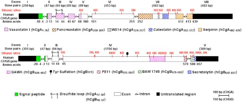
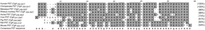
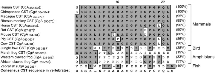
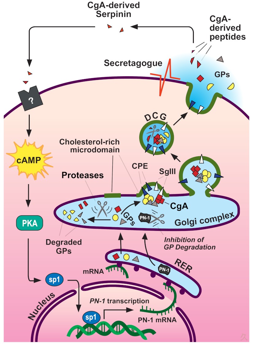
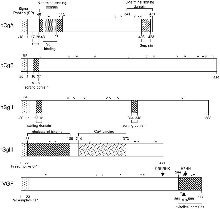
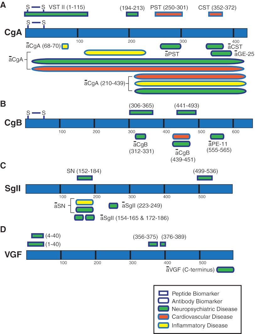
The chromogranins (chromogranin A and chromogranin B), secretogranins (secretogranin II and secretogranin III), and additional related proteins (7B2, NESP55, proSAAS, and VGF) that together comprise the granin family subserve essential roles in the regulated secretory pathway that is responsible for controlled delivery of peptides, hormones, neurotransmitters, and growth factors. Here we review the structure and function of granins and granin-derived peptides and expansive new genetic evidence, including recent single-nucleotide polymorphism mapping, genomic sequence comparisons, and analysis of transgenic and knockout mice, which together support an important and evolutionarily conserved role for these proteins in large dense-core vesicle biogenesis and regulated secretion. Recent data further indicate that their processed peptides function prominently in metabolic and glucose homeostasis, emotional behavior, pain pathways, and blood pressure modulation, suggesting future utility of granins and granin-derived peptides as novel disease biomarkers.


Figures

Fig 1.
Fig 1.
Genomic organization of human CgA and CgB. Positions of sequences encoding conserved, biologically active CgA- and CgB-derived peptides within the human CHGA and CHGB genes, respectively, are shown.
Fig. 2.
Fig. 2.
Alignment of the PST domain of CgA. PST alignment in mammalian species was performed using the ClustalW program of MacVector version 9.0, and the percentages of homology were calculated (shown in parentheses). The most conserved amino acids are highlighted in gray. PST sequences used are human (accession number NM_001275), chimpanzee (XM_510135), macaque (AB_169793), rhesus monkey (XM_001092629), horse (NM_001081814), cow (NM_181005), pig (NM_001164005), rat (NM_021655), and mouse (NM_007693).
Fig. 3.
Fig. 3.
Alignment of the CST domain of CgA. CST alignment in vertebrate species was performed using the ClustalW program of MacVector version 9.0, and the percentages of homology were calculated (shown in parentheses). The most conserved amino acids are highlighted in gray. CST sequences used are human (accession number NM_001275), chimpanzee (XM_510135), macaque (AB_169793), rhesus monkey (XM_001092629), horse (NM_001081814), rat (NM_021655), mouse (NM_007693), pig (NM_001164005), cow (NM_181005), jungle fowl (XM_421330), and marsh frog (AF139924).
Fig. 4.
Fig. 4.
Evolutionary conservation of vertebrate and invertebrate granins. Protein sequences for SgIII (SCG3), 7B2, VGF, and proSAAS were aligned using the program ClustalW2 (422), edited with Jalview version 2.5.1 (423), and displayed using the ClustalX color scheme. Amino acid numbers shown in the alignments correspond to the entire protein coding sequences for rat SCG3 (A), the mature human 7B2 protein (B), rat VGF (C), and human proSAAS (D). Three functional domains of SCG3, which bind to cholesterol, CgA, and carboxypeptidase E (CPE), each contain regions that have been highly conserved throughout vertebrate evolution (A). For 7B2, the most highly conserved regions include the cysteine-stabilized PC2 interaction domain that is conserved even in invertebrates, and the C-terminal peptide catalytic inhibitor of PC2, which is highly conserved in vertebrates (B). Three highly conserved regions of VGF, one that includes the NERP-2 peptide and another that includes the C-terminal TLQP and AQEE peptides, are each conserved in higher and lower vertebrates (C). For proSAAS, the C-terminal PC1/3 inhibitory peptide with its LLRVKRL motif is conserved in vertebrates (D). No bird orthologs of VGF or proSAAS were identified using looser BLAST search parameters (51), querying with these short conserved domains. SgIII (SCG3) sequences are human isoform 1 (NP_037375), dog (XP_535482), cow (NP_001095567), mouse (NP_0331561), rat (NP_446308), chicken (XP_413807), Xenopus (NP_001079046), and zebrafish (NP_957051). 7B2 sequences aligned are human isoform I (accession number NP_001138229), mouse (NP_033188), rat (NP_037307), cow (NP_001039463), chimpanzee (NP_001092019), zebrafish (NP_957020), Aplysia (ABF21075), C. elegans (NP_508020), and Drosophila isoform B (NP_001014608). VGF sequences aligned are human (NP_003369), rat (NP_112259), mouse (NP_001034474), cow (XP_875466), horse (XP_001916046), macaca (NC_007860), chimpanzee (XP_519275), opossum (XP_001371271), and zebrafish (XP_001343121). ProSAAS sequences included are human (NP_037403), mouse (NP_038920), rat (NP_062152), cow (NP_001077149), orangutan (XP_002831656), and zebrafish (NP_001159601).
Fig. 5.
Fig. 5.
Model for intracellular trafficking of granule proteins and autocrine regulation of DCG biogenesis by the CgA-derived peptide serpinin in endocrine cells. Granule proteins (GPs, including granins and prohormones) are synthesized at the RER and then transported to the Golgi complex where they are sorted at the TGN into the regulated secretory pathway. Granins (e.g., CgA) and prohormones form aggregates, which bind to SgIII or carboxypeptidase E (CPE), sorting receptors that are anchored to cholesterol-sphingolipid-rich membrane microdomains at the TGN. These membrane domains bud under the driving force of the granin (CgA and CgB) aggregates to form immature granules. Specific proteolytic enzymes process the prohormones fully or the granins partially to yield biologically active peptides. The granins, the major protein in the granules, condense to form mature DCG. Excess granule proteins are degraded in the Golgi complex. Upon stimulation of the cell, DCG exocytose and release their contents. In cells expressing CgA, a C-terminal peptide, serpinin, is released and binds to a putative G protein-coupled receptor to increase the transcription and biosynthesis of protease nexin-1 (PN-1) via a cAMP-PKA-Sp1 signal transduction pathway. PN-1 inhibits GP degradation to increase GP levels, which in turn leads to more DCG formation to replenish the ones secreted.
Fig. 6.
Fig. 6.
Schematic diagram showing the structures and sorting domains of bovine (b) CgA, bovine CgB, human (h) SgII, rat (r) SgIII, and rat VGF. The sorting determinants of these granins and the binding sites referred to in the text are indicated. The arrowheads represent paired or multiple basic residues that are potential PC cleavage sites. The asterisk in the VGF structure represents a noncanonical cleavage site 553RPR555 that is cleaved to generate a number of peptides.
Fig. 7.
Fig. 7.
Utility of granin biomarkers in psychiatric, neurodegenerative, cardiovascular, and inflammatory disease. Rectangles, representing peptide fragments of CgA (panel A), CgB (panel B), SgII (panel C), and VGF (panel D) that have been used as biomarkers, are color-coded for disease: psychiatric and neurodegenerative disease (green), hypertension and cardiovascular disease (red), and inflammatory disease (yellow). Granin regions that are recognized by antibodies (labeled ā) used in biomarker studies are denoted by the similarly colored ellipses. Note that ellipses outnumber rectangles, representing the current reliance on antibody-based detection methods and that the application of these granin biomarkers to neurodegenerative and psychiatric disease is comparatively more common. There are fewer biomarker reports for NESP55 (301), proSAAS (354), SgIII (424), and 7B2 (425), so these granins are not included in the illustration. The accompanying Table 5 provides the diseases that these diagnostic biomarkers have been used for and the references describing their application. S-S represents the N-terminal domain containing the disulfide bonded Cys-Cys loop structure.

References

    1. Burgess TL, Kelly RB. 1987. Constitutive and regulated secretion of proteins. Annu Rev Cell Biol 3:243–293 - PubMed
    1. Kelly RB. 1985. Pathways of protein secretion in eukaryotes. Science 230:25–32 - PubMed
    1. Arvan P, Castle D. 1998. Sorting and storage during secretory granule biogenesis: looking backward and looking forward. Biochem J 332:593–610 - PMC - PubMed
    1. Dikeakos JD, Reudelhuber TL. 2007. Sending proteins to dense core secretory granules: still a lot to sort out. J Cell Biol 177:191–196 - PMC - PubMed
    1. Park JJ, Loh YP. 2008. How peptide hormone vesicles are transported to the secretion site for exocytosis. Mol Endocrinol 22:2583–2595 - PMC - PubMed

Publication types

MeSH terms