Review
. 2011 Dec;32(6):725-54.
doi: 10.1210/er.2011-0008. Epub 2011 Aug 23.

Immunological applications of stem cells in type 1 diabetes

Affiliations
Review

Immunological applications of stem cells in type 1 diabetes

Paolo Fiorina et al. Endocr Rev. 2011 Dec.

Abstract

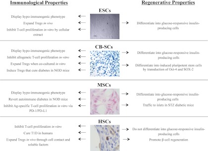
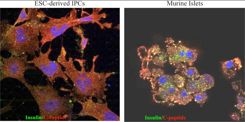
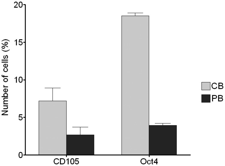
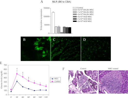
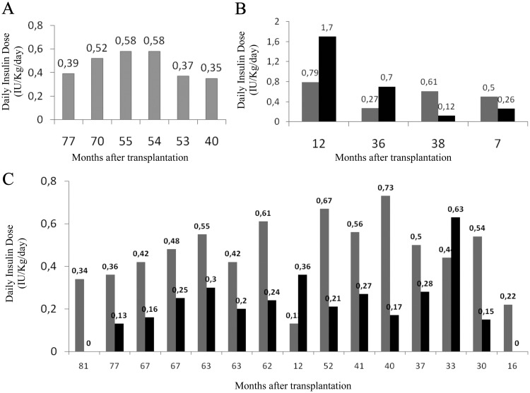
Current approaches aiming to cure type 1 diabetes (T1D) have made a negligible number of patients insulin-independent. In this review, we revisit the role of stem cell (SC)-based applications in curing T1D. The optimal therapeutic approach for T1D should ideally preserve the remaining β-cells, restore β-cell function, and protect the replaced insulin-producing cells from autoimmunity. SCs possess immunological and regenerative properties that could be harnessed to improve the treatment of T1D; indeed, SCs may reestablish peripheral tolerance toward β-cells through reshaping of the immune response and inhibition of autoreactive T-cell function. Furthermore, SC-derived insulin-producing cells are capable of engrafting and reversing hyperglycemia in mice. Bone marrow mesenchymal SCs display a hypoimmunogenic phenotype as well as a broad range of immunomodulatory capabilities, they have been shown to cure newly diabetic nonobese diabetic (NOD) mice, and they are currently undergoing evaluation in two clinical trials. Cord blood SCs have been shown to facilitate the generation of regulatory T cells, thereby reverting hyperglycemia in NOD mice. T1D patients treated with cord blood SCs also did not show any adverse reaction in the absence of major effects on glycometabolic control. Although hematopoietic SCs rarely revert hyperglycemia in NOD mice, they exhibit profound immunomodulatory properties in humans; newly hyperglycemic T1D patients have been successfully reverted to normoglycemia with autologous nonmyeloablative hematopoietic SC transplantation. Finally, embryonic SCs also offer exciting prospects because they are able to generate glucose-responsive insulin-producing cells. Easy enthusiasm should be mitigated mainly because of the potential oncogenicity of SCs.


Figures

Fig. 1.
Fig. 1.
Immunological and regenerative properties of MSCs, CB-SCs, HSCs, and ESCs. Immunoregulatory and regenerative properties are described herein for each SC line, primarily focusing on T1D-related studies. Images show MSCs (hematoxylin and eosin staining of BALB/c bone marrow-derived MSCs obtained at P4); CB-SCs (chondrogenic differentiation of CB-SCs stained with toluidine blue); HSCs (murine CD34+ cells in the wound of a diabetic mouse); and ESCs [human ES cell line (H13) culture with mouse feeder cell].
Fig. 2.
Fig. 2.
ESC-derived IPCs poorly produce insulin. ESCs were differentiated into IPCs, and cells were stained for both insulin and C-peptide. Isolated pancreatic islets were used as controls. ESC-derived IPCs displayed weak insulin staining, whereas C-peptide expression was more detectable.
Fig. 3.
Fig. 3.
The high percentage of CD105 and Oct4 positive cells in CB compared with peripheral blood (PB) confirmed the considerable naiveté of CB-SCs and their suitability for regenerative use.
Fig. 4.
Fig. 4.
Bone marrow-derived MSCs from different third-party strains (NOD or BALB/c) inhibited the MLR (A). Once injected iv into NOD mice, MSCs obtained from BALB/c mice were detectable in the pancreas at d 1 (B), in the pancreatic lymph nodes after 7 d (C), and in the spleen up to 30 d after injection (D). NOD mice injected with BALB/c MSCs showed a near-normal glucose profile upon peritoneal glucose testing as well as reduced insulitis (14 d after injection) (E and F).
Fig. 5.
Fig. 5.
Daily insulin doses in individual T1D patients before (gray bars) and after (black bars) autologous HSC transplantation. Metabolic data from different groups of patients are presented: A, patients insulin-free since transplantation (because they are continously insulin-free, only gray bars are shown); B, patients who were never insulin-free; and C, patients who were transiently insulin-free. Updated results of 25 patients (December, 2010).

References

    1. Colucci F, Cilio CM. 2010. Taming killer cells may halt diabetes progression. Nat Immunol 11:111–112 - PubMed
    1. Karges B, Kapellen T, Neu A, Hofer SE, Rohrer T, Rosenbauer J, Wolf J, Holl RW. 2010. Long-acting insulin analogs and the risk of diabetic ketoacidosis in children and adolescents with type 1 diabetes. Diabetes Care 33:1031–1033 - PMC - PubMed
    1. Nathan DM, Cleary PA, Backlund JY, Genuth SM, Lachin JM, Orchard TJ, Raskin P, Zinman B. 2005. Intensive diabetes treatment and cardiovascular disease in patients with type 1 diabetes. N Engl J Med 353:2643–2653 - PMC - PubMed
    1. Patterson CC, Dahlquist GG, Gyürüs E, Green A, Soltész G. 2009. Incidence trends for childhood type 1 diabetes in Europe during 1989–2003 and predicted new cases 2005–20: a multicentre prospective registration study. Lancet 373:2027–2033 - PubMed
    1. 2000 Retinopathy and nephropathy in patients with type 1 diabetes four years after a trial of intensive therapy The Diabetes Control and Complications Trial/Epidemiology of Diabetes Interventions and Complications Research Group. N Engl J Med [Erratum (2000) 342:1376] 342:381–389 - PMC - PubMed

Publication types

MeSH terms