. 2011 Aug;23(8):3055-69.
doi: 10.1105/tpc.111.086827. Epub 2011 Aug 23.

Elucidation of the pathway to astaxanthin in the flowers of Adonis aestivalis

Affiliations

Elucidation of the pathway to astaxanthin in the flowers of Adonis aestivalis

Francis X Cunningham Jr et al. Plant Cell. 2011 Aug.

Abstract

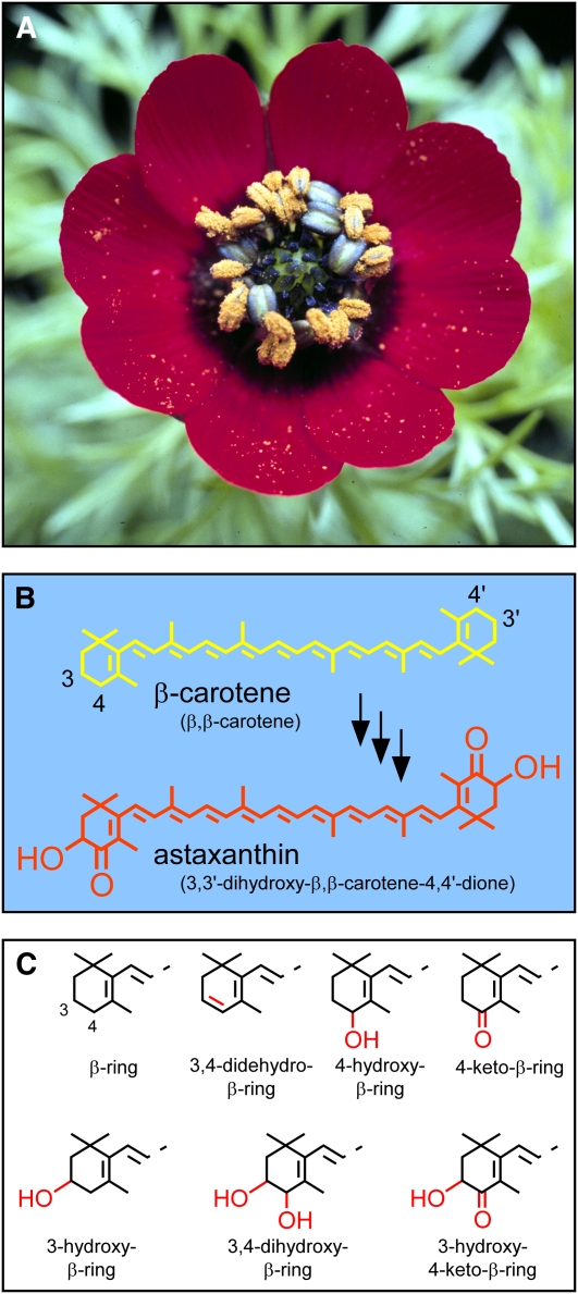
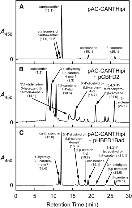
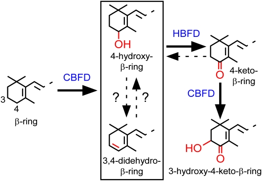
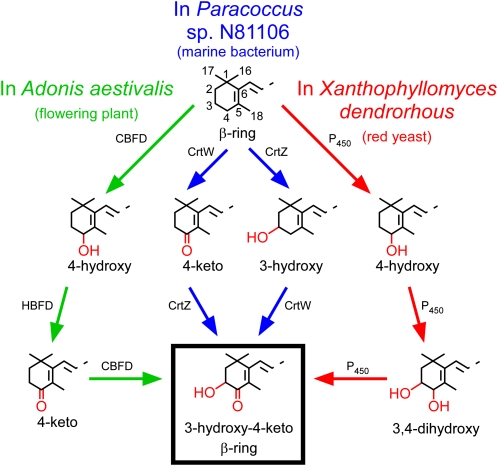
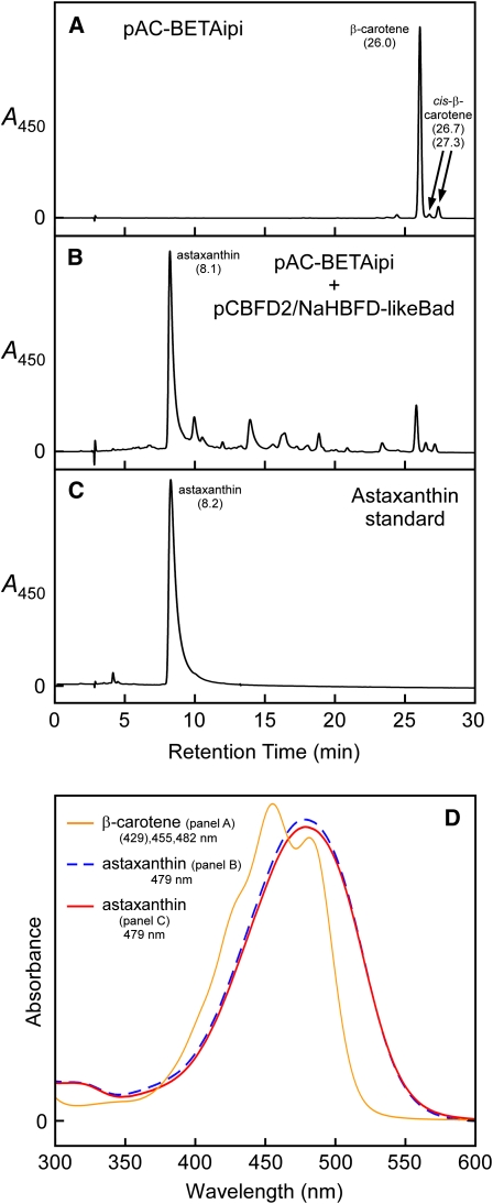
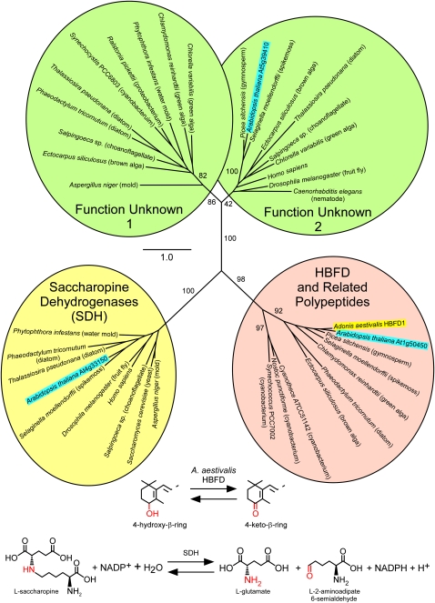
A few species in the genus Adonis are the only land plants known to produce the valuable red ketocarotenoid astaxanthin in abundance. Here, we ascertain the pathway that leads from the β-rings of β-carotene, a carotenoid ubiquitous in plants, to the 3-hydroxy-4-keto-β-rings of astaxanthin (3,3'-dihydroxy-β,β-carotene-4,4'-dione) in the blood-red flowers of Adonis aestivalis, an ornamental and medicinal plant commonly known as summer pheasant's eye. Two gene products were found to catalyze three distinct reactions, with the first and third reactions of the pathway catalyzed by the same enzyme. The pathway commences with the activation of the number 4 carbon of a β-ring in a reaction catalyzed by a carotenoid β-ring 4-dehydrogenase (CBFD), continues with the further dehydrogenation of this carbon to yield a carbonyl in a reaction catalyzed by a carotenoid 4-hydroxy-β-ring 4-dehydrogenase, and concludes with the addition of an hydroxyl group at the number 3 carbon in a reaction catalyzed by the erstwhile CBFD enzyme. The A. aestivalis pathway is both portable and robust, functioning efficiently in a simple bacterial host. Our elucidation of the pathway to astaxanthin in A. aestivalis provides enabling technology for development of a biological production process and reveals the evolutionary origin of this unusual plant pathway, one unrelated to and distinctly different from those used by bacteria, green algae, and fungi to synthesize astaxanthin.


Figures

Figure 1.
Figure 1.
A. aestivalis in Flower, Chemical Structure of the Pigment Responsible for the Resplendent Red Color of the Flower Petals, and Structures of Carotenoid Rings Relevant to This Work. (A) Mature flower of A. aestivalis. (B) Structure of astaxanthin and of the carotenoid from which it is presumed to be derived, β-carotene. Colors of the structures approximate the colors of the carotenoids they represent. Semisystematic names for the carotenoids (Weedon and Moss, 1995) are in parentheses. Numbering of the carbon atoms is indicated for two positions of interest in the two β-rings of β-carotene. (C) Structures of carotenoid rings relevant to this work. Carbon numbering for two positions of interest is indicated for the unmodified β-ring (top left). Features of interest are highlighted in red.
Figure 2.
Figure 2.
Color Complementation Screening of an A. aestivalis cDNA Library Enabled the Identification of cDNAs That Encode the Enzymes Needed to Convert β-Carotene into Astaxanthin. (A) An A. aestivalis cDNA library was introduced into a strain of E. coli that had been engineered to produce β-carotene (cells contained plasmid pAC-BETA). Cultures inoculated with the rare orange colonies (e.g., that indicated with an arrow) selected from among a multitude of yellow colonies were found to produce a complex mixture of carotenoids (lane 3 of [C]) with 4-hydroxy and/or 3,4-didehydro-β-rings (Cunningham and Gantt, 2005). (B) A second screening of the A. aestivalis cDNA library was performed in E. coli engineered to produce those pigments found in orange colonies selected in the initial screen (cells contained plasmid pAC-BETA-CBFD1/2, constructed by inserting the two cDNAs recovered from library plasmids selected in the initial screen, cbfd1 and cbfd2, into pAC-BETA). Colonies a darker orange to red in color (e.g., indicated with an arrow) were selected for analysis. (C) Reverse-phase TLC separation of pigments extracted from E. coli containing the plasmids listed below (lanes 1 to 4) and of a synthetic astaxanthin standard (lane 5). Lane 1, pAC-BETA + pHPK (contains an H. pluvialis cDNA that encodes an enzyme that converts β-carotene into echinenone and canthaxanthin); lane 2, pAC-BETA; lane 3, pAC-BETA and pCBFD1 (a cDNA library plasmid recovered from the orange colony indicated with an arrow in [A]); lane 4, pAC-BETA-CBFD1/2 and pHBFD1 (a cDNA library plasmid recovered from that colony indicated with an arrow in [B]); lane 5, astaxanthin standard. Note: The apparent colors of carotenoids on TLC plates are very much affected by the concentrations of the pigments, with higher concentrations of yellow carotenoids appearing more orange to red in color.
Figure 3.
Figure 3.
The Products of A. aestivalis cDNAs cbfd1 and hbfd1 Together Catalyze a Near Complete Conversion of β-Carotene into Astaxanthin in Cells of E. coli, with the Initial Reaction Catalyzed by the Product of cbfd1. Shown are HPLC elution profiles for extracts of E. coli cultures wherein cells contained the following plasmids: (A) pAC-BETAipi; (B) pAC-BETAipi and pCBFD1; (C) pAC-BETAipi and pHBFD1Bad; (D) pAC-BETAipi and pCBFD1/HBFD1Bad. (E) displays an HPLC elution profile for a synthetic astaxanthin standard. Absorption spectra for the peaks in (A), (D), and (E) are displayed in Supplemental Figure 6 online. Absorption spectra for the peaks in (B) are shown in Supplemental Figure 7 online. Arabinose (0.05% [w/v]) was included in the growth medium of cultures wherein cells contained pHBFD1Bad or pCBFD1/HBFD1Bad to induce production of the HBFD1 polypeptide. Numbers in parentheses below carotenoid names are HPLC retention times in minutes.
Figure 4.
Figure 4.
Time-Course Studies of the Conversion of Carotenoid β-Rings into 4-Hydroxy-β-Rings and 3-Hydroxy-4-Keto-β-Rings, as Catalyzed by the Enzymes of A. aestivalis in Cells of E. coli. (A) Time-course study for an E. coli culture wherein cells contained plasmids pAC-BETA and pCBFD2Bad. (B) Time-course study wherein cells contained pAC-BETA and pCBFD2Bad/HBFD1. Cultures were grown overnight on a rotary shaker at 28°C, diphenylamine (200 μM) was then added to block further synthesis of colored carotenoids (Cunningham and Gantt, 2007), and arabinose (0.2% [w/v]) was added 2 h later (at time = 0) to induce production of the polypeptide encoded by the A. aestivalis cbfd2 cDNA. Samples were taken at the indicated times for analysis by HPLC (see Supplemental Figure 8 online), the fraction of the total integrated area (detector set to 480 nm) was ascertained for individual peaks, and the data were plotted as a function of time after the addition of arabinose. An abbreviated scale was used for the y axis in (A) to more clearly illustrate changes in the amounts of the various intermediates over time. Accordingly, the amount of β-carotene at each time point was multiplied by 0.33 to keep on scale.
Figure 5.
Figure 5.
The Product of the A. aestivalis cbfd2 cDNA, Earlier Shown to Add a Hydroxyl to the Number 4 Carbon of Carotenoid β-Rings (Cunningham and Gantt, 2005), Adds a Hydroxyl to the Number 3 Carbon of 4-Keto-β-Rings. Shown are HPLC elution profiles for extracts of E. coli cultures wherein cells contained the following: (A) pAC-CANTHipi; (B) pAC-CANTHipi and pCBFD2; and (C) pAC-CANTHipi and pHBFD1Bad. Absorption spectra for the peaks in (A) are displayed in Supplemental Figure 6 online. Absorption spectra for the peaks in (B) and (C) are shown in Supplemental Figure 7 online. Arabinose (0.05% [w/v]) was included in the growth medium of the E. coli culture wherein cells contained pHBFD1Bad. Numbers in parentheses are HPLC retention times in minutes. Those peak identifications followed by question marks in (B) and (C) are speculative: They are consistent with absorption spectra, HPLC retention times, and the known catalytic capabilities of the carotenoid pathway enzymes that are present in the E. coli cells, but the requisite standards were not available for comparison.
Figure 6.
Figure 6.
The Pathway from Carotenoid β-Rings to 3-Hydroxy-4-Keto-β-Rings as Catalyzed by the Products of the cbfd and hbfd cDNAs of A. aestivalis in Cells of E. coli. The specific substrate for the enzyme encoded by the A. aestivalis hbfd cDNAs, whether a 3,4-didehydro-β-ring or a 4-hydroxy-β-ring, and the interconvertibility of these two rings remain uncertain. [See online article for color version of this figure.]
Figure 7.
Figure 7.
Distinctly Different Biosynthetic Pathways Serve to Convert β-Rings into 3-Hydroxy-4-Keto-β-Rings in Carotenoids of the Flowering Plant A. aestivalis, the Marine Bacterium Paracoccus sp N81106, and the Red Yeast X. dendrorhous. The pathway in Paracoccus sp N81106 (previously known as Agrobacterium aurantiacum) was ascertained by Misawa et al. (1995b). The pathway in X. dendrorhous (also known as Phaffia rhodozyma) has not been definitively established but is thought to operate using a single cytochrome P450 enzyme (with a cytochrome P450 reductase also needed to donate electrons; Alcaíno et al., 2008; Ukibe et al., 2009) to sequentially oxidize the number 3 and number 4 carbons of the two β-rings of β-carotene (Ojima et al., 2006; Álvarez et al., 2006; Martín et al., 2008). Note that the biosynthesis of carotenoids with 3-hydroxy-4-keto-β-rings in the green alga H. pluvialis entails the use of an enzyme similar in amino acid sequence to the CrtW of Paracoccus sp N81106 to catalyze the addition of the carbonyl (Kajiwara et al., 1995; Lotan and Hirschberg, 1995), but hydroxylation of the number 3 carbon may be catalyzed by a cytochrome P450 enzyme (Schoefs et al., 2001) rather than a CrtZ (or the related CHYb) enzyme. Note also that certain bacteria and cyanobacteria employ a fourth way, using an enzyme referred to as CrtO, to add a carbonyl to the number 4 carbon of carotenoid β-rings (Fernández-González et al., 1997; Tao and Cheng, 2004). A fourth way is available, as well, to add a 3-hydroxyl group to carotenoid β-rings: A carotenoid β-ring 3-hydroxylase enzyme of a type referred to as CrtR is present in certain cyanobacteria (Masamoto et al., 1998). CBFD, the carotenoid β-ring 4-dehydrogenase enzyme encoded by the A. aestivalis cbfd1 and cbfd2 cDNAs; HBFD, the carotenoid 4-hydroxy-β-ring 4-dehydrogenase enzyme encoded by the A. aestivalis hbfd1 and hbfd2 cDNAs.
Figure 8.
Figure 8.
Maximum Likelihood Trees for the CBFD and HBFD Enzymes of A. aestivalis and Related Polypeptides Encoded by cDNAs and Genes of Other Plants and Green Algae. (A) Tree for plant and green algal carotenoid β-ring 3-hydroxylase enzymes of the membrane-integral, diiron, nonheme oxygenase type (CHYb-type) together with the A. aestivalis CBFD1 and CBFD2 polypeptides. CBFD1 and CBFD2 are highlighted with a red background. The A. aestivalis CHYb is enclosed in a red box. The amino acid sequences of CBFD1 and CBFD2 are ~90% identical to each other and ~58 to 60% identical overall to the A. aestivalis CHYb. (B) Tree for the A. aestivalis HBFD1 and HBFD2 enzymes together with related polypeptides encoded by cDNAs and genes of other plants and green algae. HBFD1 and HBFD2 are highlighted with a red background, as is a N. advena polypeptide also shown to have 4-hydroxy-β-ring 4-dehydrogenase activity (Figure 9). Green algal sequences served as an outgroup for each tree. One thousand bootstrap trials were conducted, with bootstrap values >50% indicated. Alignments used to construct these trees are shown in Supplemental Figures 3 (for the CBFD and CHYb enzymes) and 4 (for HBFD and related polypeptides) online and are available in FASTA format as Supplemental Data Sets 1 and 2 online, respectively.
Figure 9.
Figure 9.
An N. advena cDNA That Encodes a Polypeptide (NaHBFD-Like) Similar in Amino Acid Sequence to the Products of the A. aestivalis hbfd1 and hbfd2 cDNAs, When Combined with the A. aestivalis cbfd2 cDNA, Leads to the Conversion of β-Carotene into Astaxanthin in Cells of E. coli. Shown are HPLC elution profiles for extracts of E. coli cultures wherein cells contained the following plasmids: (A) pAC-BETAipi and (B) pAC-BETAipi and pCBFD2/NaHBFD-likeBad. (C) displays an HPLC elution profile for a synthetic astaxanthin standard, and (D) shows absorption spectra for the major peaks of (A) to (C). Note: The N. advena cDNA was fused, in frame, to the arabinose-inducible araBAD promoter to ensure high-level expression. Arabinose (0.2% [w/v]) was included in the growth medium for that culture containing pCBFD2/NaHBFD-likeBad. Numbers in parentheses below carotenoid names are HPLC retention times in minutes. [See online article for color version of this figure.]
Figure 10.
Figure 10.
Maximum Likelihood Tree for Selected Members of the Extended Saccharopine Dehydrogenase Family of Enzymes. Each of the four clades encompasses polypeptides from a great diversity of species. One clade contains polypeptides that, in several cases at least, have been shown to function as SDHs (saccharopine dehydrogenase [NADP(+), l-glutamate-forming]; EC 1.5.1.10). Note: This enzyme, often referred to as saccharopine reductase, is unrelated to the l-lysine–forming saccharopine dehydrogenase (EC 1.5.1.7). A second clade contains the A. aestivalis HBFD1 enzyme and related polypeptides of unknown function from Arabidopsis and other plants, algae, and cyanobacteria. Two additional clades, labeled “Function Unknown 1” and “Function Unknown 2,” contain polypeptides whose functions have not been ascertained. The A. aestivalis HBFD1 is highlighted with a yellow background. Three Arabidopsis polypeptides are each highlighted with a bright-blue background. The reactions catalyzed by SDH and by the A. aestivalis HBFD are illustrated below the tree. Like that catalyzed by SDH, the reaction catalyzed by HBFD probably requires the participation of NADP+, but this has not yet been demonstrated. One thousand bootstrap trials were conducted, with bootstrap support values indicated for the major branches. The alignment used to construct this tree is shown in Supplemental Figure 5 online and is available in FASTA format as Supplemental Data Set 3 online.

References

    1. Alcaíno J., Barahona S., Carmona M., Lozano C., Marcoleta A., Niklitschek M., Sepúlveda D., Baeza M., Cifuentes V. (2008). Cloning of the cytochrome p450 reductase (crtR) gene and its involvement in the astaxanthin biosynthesis of Xanthophyllomyces dendrorhous. BMC Microbiol. 8: 169 - PMC - PubMed
    1. Álvarez V., Rodríguez-Sáiz M., de la Fuente J.L., Gudiña E.J., Godio R.P., Martín J.F., Barredo J.L. (2006). The crtS gene of Xanthophyllomyces dendrorhous encodes a novel cytochrome-P450 hydroxylase involved in the conversion of β-carotene into astaxanthin and other xanthophylls. Fungal Genet. Biol. 43: 261–272 - PubMed
    1. Andi B., Cook P.F., West A.H. (2006). Crystal structure of the his-tagged saccharopine reductase from Saccharomyces cerevisiae at 1.7-Å resolution. Cell Biochem. Biophys. 46: 17–26 - PubMed
    1. Bauernfeind J.C., Adams C.R., Marusich W.L. (1981). Carotenes and other vitamin A precursors in animal feed. In Carotenoids as Colorants and Vitamin A Precursors: Technological and Nutritional Applications, Bauernfeind J.C., (New York: Academic Press; ), pp. 563–743
    1. Broadwater J.A., Whittle E., Shanklin J. (2002). Desaturation and hydroxylation. Residues 148 and 324 of Arabidopsis FAD2, in addition to substrate chain length, exert a major influence in partitioning of catalytic specificity. J. Biol. Chem. 277: 15613–15620 - PubMed

Publication types

MeSH terms

Associated data

LinkOut - more resources