. 2011 Sep 1;10(17):2994-3002.
doi: 10.4161/cc.10.17.17118. Epub 2011 Sep 1.

Crosstalk between MYCN and MDM2-p53 signal pathways regulates tumor cell growth and apoptosis in neuroblastoma

Affiliations

Crosstalk between MYCN and MDM2-p53 signal pathways regulates tumor cell growth and apoptosis in neuroblastoma

Jing He et al. Cell Cycle. .

Abstract

Previous studies show that the MYCN and MDM2-p53 signal pathways are mutually regulated: MYCN stimulates MDM2 and p53 transcription, whereas MDM2 stabilizes MYCN mRNA and induces its translation. Herein, we report that the interaction between MDM2 and MYCN plays a critical role in MYCN-amplified neuroblastoma tumor cell growth and survival. Distinct from the known role that MDM2 has in regulating tumor promotion in non-MYCN-amplified neuroblastoma, in which MDM2 inhibits p53, we found that MDM2 stimulated tumor growth in MYCN-amplified neuroblastoma in a p53-independent manner. In MYCN-amplified neuroblastoma cells, enforced expression of MDM2 further enhanced MYCN expression, yet no p53 inhibition was observed by MDM2 due to upregulation of MYCN that stimulated p53 transcription. Similarly, p53 expression remained unchanged in MDM2-silenced MYCN-amplified neuroblastoma cells because MDM2 inhibition resulted in a downregulation of MYCN that decreased p53 transcription, although the MDM2-mediated degradation of p53 was reduced. Also, we found that the enforced overexpression of MDM2, or conversely, the inhibition of overexpressed endogenous MDM2, led to either a remarkable increase or decrease in tumor growth, respectively, in MYCN-amplified neuroblastoma (even though no p53 function was involved). These results suggest that p53 that is reciprocally regulated by MDM2 and MYCN is dispensable for suppression of MYCN-amplified neuroblastoma, and that the direct interaction between MDM2 and MYCN may contribute significantly to MYCN-amplified neuroblastoma growth and disease progression.


Figures

Figure 1
Figure 1
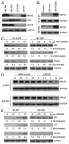
The different effects of MDM2 on both the regulation of p53 expression and its function, in neuroblastoma cells with and without MYCN amplification. (A) Western blot assay for protein expression of MDM2, p53 and MYCN in four cultured neuroblastoma cell lines. (B) Enforced overexpression of MDM2 in SK-N-SH and NB-1691 cells by transfection of the expression plasmid CMV-MDM2, as detected by western blot analysis. (C) Expression of p53 and its down-stream target genes p21 and PUMA in MDM2-transfected cells, as detected by western blot assay. Labels under bands in the blot represent the protein levels after normalization to GAPDH, as compared with samples (0 was defined as 1 unit). (D) Inhibition of endogenous MDM2 by siRNA in SH-EP 1 and NB-1691 cells. Cells were transfected with different doses of the siMDM2 and siRNA control, as indicated. After a 24-h transfection, MDM2 expression was detected by western blot. (E) The expression of p53, p21 and PUMA in two MDM2-silenced cell lines with different MYCN expression, as detected by western blot assay.
Figure 2
Figure 2
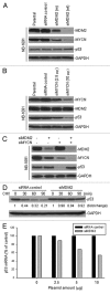
The effect of inhibition of MDM2 and MYCN on the regulation of p53 in MYCN-amplified/MDM2-overexpressed neuroblastoma cells. (A) NB-1691 cells were transfected with either siMDM2 (wt), siMDM2 (m) or siRNA control, and the expression of MDM2, MYCN and p53 was detected by western blot assay. (B) NB-1691 cells were transfected with different amount of siMYCN and expression levels of MYCN, MDM2 and p53 were detected by western blot. (C) Expression of indicated proteins as detected by western blot, in NB-1691 cells that were transfected with siMDM2, in the presence or absence of co-transfection with siMYCN. (D) p53 protein turnover in NB-1691 cells transfected with siMDM2 and control siRNA. siRNA-transfected cells were treated with the protein synthesis inhibitor cyclohexamide (CHX). At selected times indicated after CHX, cell lysates were prepared and analyzed by western blot assay. (E) p53 mRNA expression in NB-1691 transfected with siMDM2 and siRNA control was detected by quantitative RT-PCR.
Figure 3
Figure 3
The effect of MDM2 and MYCN modification on the regulation of p53 in neuroblastoma cells. (A) The MYCN-amplified NB-1643 cells were transfected with different amounts of MDM2 expression plasmid, as indicated. The expression levels of transfected MDM2, endogenous MYCN and p53 were tested by western blot assay. (B) Expression of MDM2, MYCN and p53 in NB-1643 cells transfected with MDM2, in the presence or absence of MYCN knockdown by transfection of siMYCN. (C) The SK-N-SH cells without MYCN amplification were transfected with a MYCN expression plasmid. Expression levels of the transfected MYCN, endogenous MDM2 and p53 were detected by western blot. (D) Effect of MDM2 inhibition by siRNA on regulation of p53 and ectopic MYCN in SHEP-Tet/21N cells in the presence and absence of tetracycline (Tet). Cells were cultured in medium with or without 1 µg/ml Tet and transfected with different amounts of siMDM2 for 24 h. The expression of proteins as indicated was detected by western blot.
Figure 4
Figure 4
Different effects of MDM2 inhibition on the regulation of cell growth and apoptosis in MYCN-amplified and non-MYCN-amplified neuroblastoma. (A) Clonogenic assay of SH-EP 1 and NB-1691 cells transfected with siMDM2 (wt), siMDM2 (m) and siRNA control. Cells (1 × 10⁴) were seeded in soft agarose and cultured for 2–3 weeks. Colonies were counted under phase contrast microscopy. Data represent the mean of three independent experiments; bars ± SD (B) A cell cycle analysis in SH-EP 1 and NB-1691 cells, performed 24 h after transfection with siMDM2 and siRNA control. (C) Representative histograph of flow cytometry for detection of apoptosis in SH-EP 1 and NB-1691 cells after 24 h siMDM2 transfection with different amounts of plasmid, as indicated. Apoptotic cells were detected by annexin-V staining followed by flow cytometry. (D) The effects of MDM2 inhibition by siRNA on the sensitivity of SH-EP -1 and NB-1691 neuroblastoma cell lines to etoposide-induced apoptosis. The siMDM2 and siRNA control cells were treated with 10 µM etoposide for the indicated time, and then apoptotic cells were detected by flow cytometry, *p < 0.01.
Figure 5
Figure 5
Effects of MDM2 modulation on the cell growth of MYCN-overexpressed neuroblastoma. (A) Clonogenic assay of NB-1643 cells that were stably transfected with MDM2, as compared with parental cells in the presence (siMYCN) or absence (siRNA control) of MYCN siRNA. (B) Comparison of the effect of MDM2 inhibition on growth of cells with and without conditional MYCN expression. SHEP -Tet/21N cells in cultures with or without tetracycline (Tet) were similarly transfected with siMDM2 and control siRNA, and clonogenic assays were performed as in (A), *p < 0.01. (C) Stable suppression of MDM2 in p53-null LA1-55N cells by transfection of siMDM2. MDM2 and MYCN expression in parental LA1-55N cells and LA1-55N that were transfected with the siRNA control, siMDM2 (m), and siMDM2 (wt), as detected by western blot. (D) Clonogenic assay of LA1-55N cells transfected with siRNA control, siMDM2 (clone wt 2), and siMDM2 (m), or the untransfected parental cell line. Cell culture and colony counts were performed as in (A).

Similar articles

Cited by

References

    1. Bowen KA, Chung DH. Recent advances in neuroblastoma. Curr Opin Pediatr. 2009;21:350–356. doi: 10.1097/MOP.0b013e32832b1240. - DOI - PubMed
    1. Mueller S, Matthay KK. Neuroblastoma: biology and staging. Curr Oncol Rep. 2009;11:431–438. doi: 10.1007/s11912-009-0059-6. - DOI - PubMed
    1. Maris JM, Matthay KK. Molecular biology of neuroblastoma. J Clin Oncol. 1999;17:2264–2279. - PubMed
    1. Brodeur GM, Seeger RC, Schwab M, Varmus HE, Bishop JM. Amplification of N-myc in untreated human neuroblastomas correlates with advanced disease stage. Science. 1984;224:1121–1124. doi: 10.1126/science.6719137. - DOI - PubMed
    1. Seeger RC, Brodeur GM, Sather H, Dalton A, Siegel SE, Wong KY, et al. Association of multiple copies of the N-myc oncogene with rapid progression of neuroblastomas. N Engl J Med. 1985;313:1111–1116. doi: 10.1056/NEJM198510313131802. - DOI - PubMed

Publication types

MeSH terms