. 2011 Sep 27;105(7):996-1001.
doi: 10.1038/bjc.2011.330. Epub 2011 Aug 23.

Upregulation of cancer-associated myofibroblasts by TGF-β from scirrhous gastric carcinoma cells

Affiliations

Upregulation of cancer-associated myofibroblasts by TGF-β from scirrhous gastric carcinoma cells

Y Fuyuhiro et al. Br J Cancer. .

Abstract

Background: Myofibroblasts in the cancer microenvironment have recently been implicated in tumour growth and metastasis of gastric cancer. However, the mechanisms responsible for the regulation of myofibroblasts in cancer-associated fibroblasts (CAFs) remain unclear. This study was performed to clarify the mechanisms for regulation of myofibroblasts in gastric cancer microenvironment.

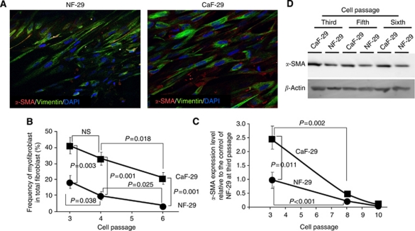
Methods: Two CAFs (CaF-29 and CaF-33) from the tumoural gastric wall and a normal fibroblast (NF-29) from the nontumoural gastric wall, 4 human gastric cancer cell lines from scirrhous gastric cancer (OCUM-2MD3 and OCUM-12), and non-scirrhous gastric cancer (MKN-45 and MKN-74) were used. Immunofluorescence microscopy by triple-immunofluorescence labelling (α-SMA, vimentin, and DAPI) was performed to determine the presence of α-SMA-positive myofibroblasts. Real-time RT-PCR was performed to examine α-SMA mRNA expression.

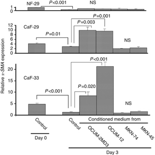
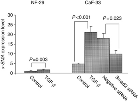
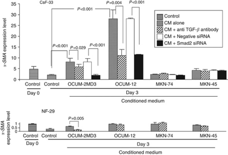
Results: Immunofluorescence microscopy showed that the frequency of myofibroblasts in CaF-29 was greater than that in NF-29. The number of myofibroblasts in gastric fibroblasts gradually decreased with serial passages. Transforming growth factor-β (TGF-β) significantly increased the α-SMA expression level of CAFs. Conditioned medium from OCUM-2MD3 or OCUM-12 cells upregulated the α-SMA expression level of CAFs, but that from MKN-45 or MKN-74 cells did not. The α-SMA upregulation effect of conditioned medium from OCUM-2MD3 or OCUM-12 cells was significantly decreased by an anti-TGF-β antibody or Smad2 siRNA.

Conclusion: Transforming growth factor-β from scirrhous gastric carcinoma cells upregulates the number of myofibroblasts in CAFs.


Conflict of interest statement

The authors declare no conflict of interest.

Figures

Figure 1
Figure 1
The α-smooth muscle actin (α-SMA) expression in fibroblasts. (A) Immunofluorescence of NF-29 fibroblasts and CaF-29 fibroblasts. Fibroblasts were stained with α-SMA (red), vimentin (green), and cell nuclei were stained with DAPI (blue). The percentage of myofibroblasts accompanying cancer-associated fibroblasts, CaF-29, from gastric tumour lesions was higher than that from normal fibroblasts, NF-29, derived from normal gastric tissue. (B) The proportion of myofibroblasts in the primary culture. The percentage of α-SMA-positive myofibroblast cells was determined in 10 random fields. The percentage of α-SMA-positive myofibroblasts cells of NF-29 (•) and CaF-29 (▪) at the third passage were 42% and 18%, respectively. At the fourth or sixth passage, the myofibroblast contents of both NF-29 and CaF-29 fibroblast cultures were lower than that at the third passage. (C) The expression level of α-SMA mRNA in the primary culture. The α-SMA expression level of CaF-29 (•) at the third passage was 2.5 relative to the α-SMA expression level of NF-29 (▪) as the control. The α-SMA expression level of both NF-29 and CaF-29 at the eighth or tenth passage was under 0.3 relative to the control of NF-29 at the third passage. (D) Western blot analysis. The α-SMA expression level in CaF-29 was higher than that of NF-29 at each passage. The α-SMA expression level in CaF-29 at the third passage was high in comparison with that at fifth or sixth passage.
Figure 2
Figure 2
Effect of conditioned medium from gastric cancer cells on the α-SMA expression of fibroblast. The α-SMA expression levels of cancer-associated fibroblasts CaF-29 and CaF-33 were significantly increased by conditioned medium from scirrhous gastric cancer cells OCUM-2MD3 and OCUM-12, but not by conditioned medium from non-scirrhous gastric cancer cells MKN-45 and MKN-74. The α-SMA expression of normal NF-29 fibroblasts was not increased by the addition of conditioned medium from any gastric cancer cells. The graph depicts expression levels relative to control NF-29 fibroblasts at day 0.
Figure 3
Figure 3
Effect of TGF-β or Smad2 siRNA on α-SMA expression of fibroblasts. The TGF-β (10 ng ml–1) increased the α-SMA expression level of gastric fibroblasts. The upregulation of α-SMA expression level by TGF-β was significantly (P=0.023) decreased by Smad2 siRNA (30 nM) in CaF-33 cells. The graph depicts expression levels relative to control NF-29.
Figure 4
Figure 4
Effect of anti-TGF-β antibody or Smad2 siRNA on α-SMA expression of fibroblasts. The conditioned medium (CM) from OCUM-2MD3 and OCUM-12 cells significantly increased the α-SMA expression level of cancer-associated fibroblast CaF-33 cells. The α-SMA expression level of CaF-33 with the addition of CM of OCUM-2MD3 and OCUM-12 was significantly decreased by anti-TGF-β antibody or Smad2 siRNA (30 nM). In contrast, CM from MKN-45 and MKN-74 did not affect the α-SMA expression level of CaF-33. The graph depicts expression levels relative to control NF-29 fibroblasts at day 0.

Similar articles

Cited by

References

    1. Brenmoehl J, Miller SN, Hofmann C, Vogl D, Falk W, Scholmerich J, Rogler G (2009) Transforming growth factor-beta 1 induces intestinal myofibroblast differentiation and modulates their migration. World J Gastroenterol 15: 1431–1442 - PMC - PubMed
    1. Casey TM, Eneman J, Crocker A, White J, Tessitore J, Stanley M, Harlow S, Bunn JY, Weaver D, Muss H, Plaut K (2008) Cancer associated fibroblasts stimulated by transforming growth factor beta1 (TGF-beta 1) increase invasion rate of tumor cells: a population study. Breast Cancer Res Treat 110: 39–49 - PubMed
    1. De Wever O, Demetter P, Mareel M, Bracke M (2008) Stromal myofibroblasts are drivers of invasive cancer growth. Int J Cancer 123: 2229–2238 - PubMed
    1. De Wever O, Nguyen QD, Van Hoorde L, Bracke M, Bruyneel E, Gespach C, Mareel M (2004) Tenascin-C and SF/HGF produced by myofibroblasts in vitro provide convergent pro-invasive signals to human colon cancer cells through RhoA and Rac. FASEB J 18: 1016–1018 - PubMed
    1. Desmouliere A, Guyot C, Gabbiani G (2004) The stroma reaction myofibroblast: a key player in the control of tumor cell behavior. Int J Dev Biol 48: 509–517 - PubMed

Publication types

MeSH terms