

miR-21 and miR-155 are associated with mitotic activity and lesion depth of borderline melanocytic lesions
- PMID: 21863027
- PMCID: PMC3185929
- DOI: 10.1038/bjc.2011.288
miR-21 and miR-155 are associated with mitotic activity and lesion depth of borderline melanocytic lesions
Abstract
Background: Expression of microRNAs (miRs) has been shown to be altered in many solid tumours and is being explored in melanoma. The malignant potential of some melanocytic lesions is difficult to predict. We hypothesised that characterisation of miR expression in borderline melanocytic proliferations would lead to the identification of a molecular profile that could be used with known prognostic factors to differentiate lesions with high malignant potential.
Methods: The miR expression profile of melanocytic lesions (benign naevi, malignant melanoma and borderline melanocytic tumours) was evaluated by real-time PCR.
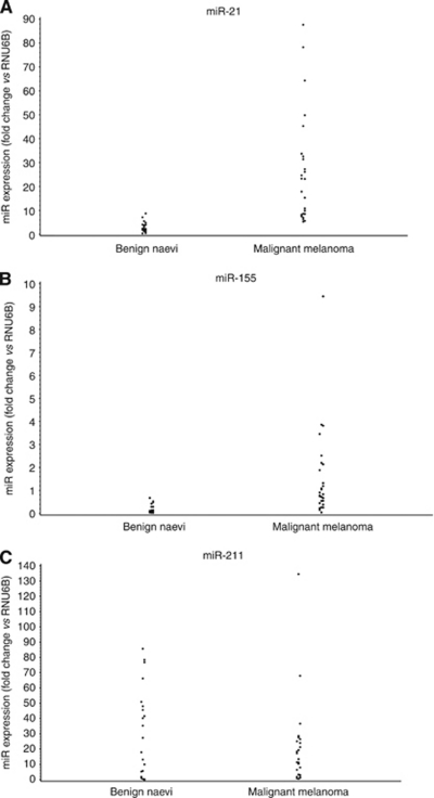
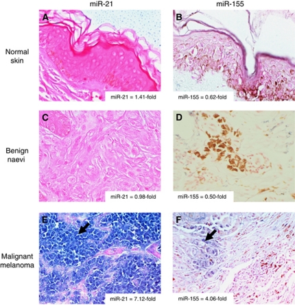
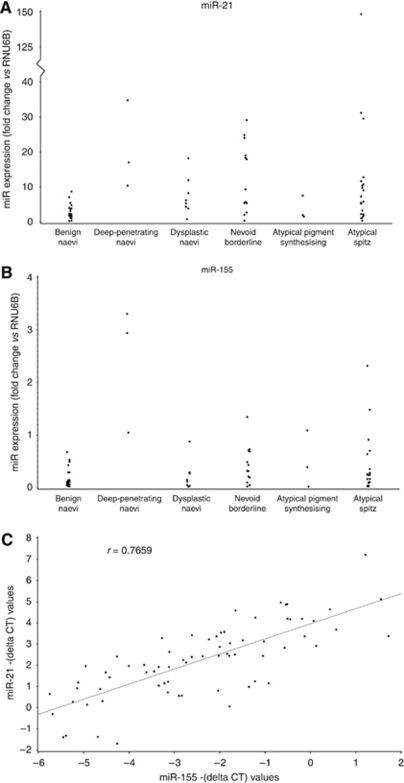
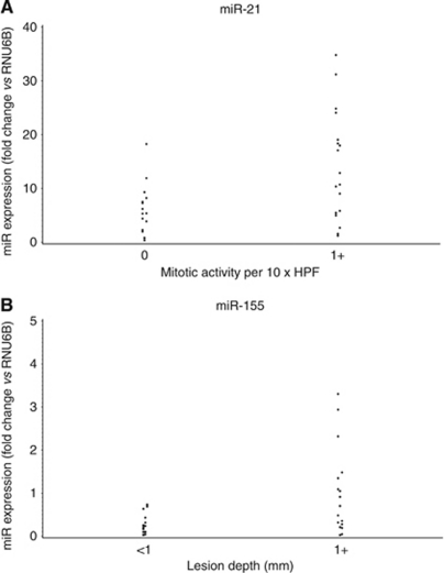
Results: PCR analysis revealed primary cutaneous melanomas had an 8.6-fold overexpression of miR-21 and a 7.5-fold overexpression of miR-155 compared with benign naevi (P<0.0001). In situ hybridisation confirmed these results. miR-21 and miR-155 were significantly overexpressed within borderline lesions (P=0.0011 and P=0.0048, respectively). When borderline lesions were categorised by mitotic activity and Breslow thickness, miR-21 was associated with mitotic activity and miR-155 was associated with thickness (P<0.025). Among 14 patients with borderline lesions who underwent sentinel lymph node biopsy (SLNB), positive SLNB was associated with increased miR-21 and miR-155 in the primary lesion compared with lesions with a negative SLNB.
Conclusion: MicroRNA expression profiles can be used to characterise atypical melanocytic lesions.
Figures
References
-
- Asangani IA, Rasheed SA, Nikolova DA, Leupold JH, Colburn NH, Post S, Allgayer H (2008) MicroRNA-21 (miR-21) post-transcriptionally downregulates tumor suppressor Pdcd4 and stimulates invasion, intravasation and metastasis in colorectal cancer. Oncogene 27: 2128–2136 - PubMed
-
- Balch CM, Soong SJ, Gershenwald JE, Thompson JF, Reintgen DS, Cascinelli N, Urist M, McMasters KM, Ross MI, Kirkwood JM, Atkins MB, Thompson JA, Coit DG, Byrd D, Desmond R, Zhang Y, Liu PY, Lyman GH, Morabito A (2001) Prognostic factors analysis of 17,600 melanoma patients: validation of the American Joint Committee on Cancer melanoma staging system. J Clin Oncol 19: 3622–3634 - PubMed
-
- Barnhill RL, Cerroni L, Cook M, Elder DE, Kerl H, LeBoit PE, McCarthy SW, Mihm MC, Mooi WJ, Piepkorn MW, Prieto VG, Scolyer RA (2010) State of the art, nomenclature, and points of consensus and controversy concerning benign melanocytic lesions: outcome of an international workshop. Adv Anat Pathol 17: 73–90 - PubMed
-
- Barnhill RL, Piepkorn MW, Busam KJ (2003) Pathology of Melanocytic Nevi and Malignant Melanoma. Springer: New York
-
- Blower PE, Chung JH, Verducci JS, Lin S, Park JK, Dai Z, Liu CG, Schmittgen TD, Reinhold WC, Croce CM, Weinstein JN, Sadee W (2008) MicroRNAs modulate the chemosensitivity of tumor cells. Mol Cancer Ther 7: 1–9 - PubMed
Publication types
MeSH terms
Substances
Grants and funding
LinkOut - more resources
Full Text Sources
Other Literature Sources
Medical
