. 2011 Sep 27;105(7):1002-11.
doi: 10.1038/bjc.2011.303. Epub 2011 Aug 23.

A truncated form of CD9-partner 1 (CD9P-1), GS-168AT2, potently inhibits in vivo tumour-induced angiogenesis and tumour growth

Affiliations

A truncated form of CD9-partner 1 (CD9P-1), GS-168AT2, potently inhibits in vivo tumour-induced angiogenesis and tumour growth

S Colin et al. Br J Cancer. .

Abstract

Background: Tetraspanins are transmembrane proteins known to contribute to angiogenesis. CD9 partner-1 (CD9P-1/EWI-F), a glycosylated type 1 transmembrane immunoglobulin, is a member of the tetraspanin web, but its role in angiogenesis remains to be elucidated.

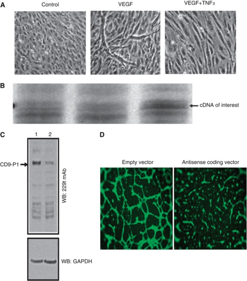
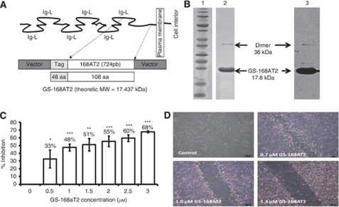
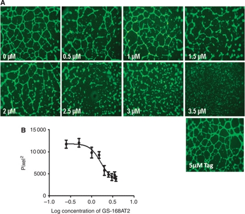
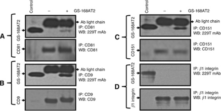
Methods: We measured the expression of CD9P-1 under angiogenic and angiostatic conditions, and the influence of its knockdown onto capillary structures formation by human endothelial cells (hECs). A truncated form of CDP-1, GS-168AT2, was produced and challenged vs hEC proliferation, migration and capillaries' formation. Its association with CD9P-1, CD9, CD81 and CD151 and the expressions of these later at hEC surface were analysed. Finally, its effects onto in vivo tumour-induced angiogenesis and tumour growth were investigated.

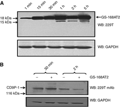
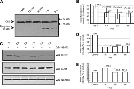
Results: Vascular endothelial growth factor (VEGF)-induced capillary tube-like formation was inhibited by tumour necrosis factor α and was associated with a rise in CD9P-1 mRNA expression (P<0.05); accordingly, knockdown of CD9P-1 inhibited VEGF-dependent in vitro angiogenesis. GS-168AT2 dose-dependently inhibited in vitro angiogenesis, hEC migration and proliferation (P<0.05). Co-precipitation experiments suggest that GS-168AT2 corresponds to the sequence by which CD9P-1 physiologically associates with CD81. GS-168AT2 induced the depletion of CD151, CD9 and CD9P-1 from hEC surface, correlating with GS-168AT2 degradation. Finally, in vivo injections of GS-168AT2 inhibited tumour-associated angiogenesis by 53.4±9.5% (P=0.03), and reduced tumour growth of Calu 6 tumour xenografts by 73.9±16.4% (P=0.007) without bodyweight loss.

Conclusion: The truncated form of CD9P-1, GS-168AT2, potently inhibits angiogenesis and cell migration by at least the downregulation of CD151 and CD9, which provides the first evidences for the central role of CD9P-1 in tumour-associated angiogenesis and tumour growth.


Figures

Figure 1
Figure 1
Angiogenesis modulation by CD9P-1 expression. (A) Human EC were incubated with culture medium alone (control), or supplemented with either VEGF (50 ng ml–1), or TNFα at antiangiogenic concentration (50 ng ml–1), for 48 h with reload of factors at 24 h. (B) Gene profiling of the differentially regulated genes in the above conditions. The cDNA of interest marked with an arrow, was identified by sequencing as the CD9-P1 gene. (C) Representative image of WB of BAEC harbouring the empty (lane 1) or CD9-P1 antisense transcript coding pci-neovector (lane 2) using 229T mAb. (D) Representative images of in vitro angiogenesis assay with BAEC harbouring the empty or CD9P-1 antisense transcript coding pci-neovector.
Figure 2
Figure 2
The truncated form of CD9-P1, GS-168AT2, inhibits dose-dependently in vitro hEC proliferation and migration. (A) Schematic representation of the structure of CD9-P1, the cloned truncated CD9-P1 domain, GS-168AT2, and the produced Tag-recombinant protein. (B) SDS–PAGE imprints (lane 1, molecular mass marker; lane 2 purified GS-168AT2) and the corresponding WB (lane 3, using 229 mAb) of the purified GS-168AT2 used for the all subsequent experiments. (C) Dose-response curve of the effects of GS168AT2 on the proliferation of hEC. Results were expressed as percentage of control±s.e. (n=4). Statistical significance between the control and the different doses of GS-168AT2 were calculated with two-tailed Student's t-test (*P<0.05; **P<0.01; ***P<0.001). (D) Representative images at 18 h of the wounded hEC monolayer incubated with either vehicle or increasing concentrations of GS-168AT2.
Figure 3
Figure 3
The truncated form of CD9P-1, GS-168AT2, inhibits dose-dependently in vitro angiogenesis. (A) Representative images showing that GS-168AT2 inhibits in vitro angiogenesis in a concentration-dependent manner, and (B) its quantification. Results were expressed as mean±s.e. (n=4).
Figure 4
Figure 4
GS-168AT2 co-precipitates with both CD9 and CD15, but poorly with CD81. Human EC were incubated with vehicle or GS-168AT2 for 2 h, washed twice with vehicle and hEC were lysed in 1% Brij 97 lysis buffer, and the indicated tetraspanins were immunoprecipitated (CD9, CD81 and CD151) by incubating the cell lysate (the protein contents of cell lysates were adjusted by Bradford protein assay) with the indicated antibody at 4 °C. The immunocomplexes were pulled down with agarose beads coated with G-protein, and the beads were washed twice with lysis buffer. The immunoprecipitates were resolved in SDS–PAGE and western blotted with the indicated antibody. GS-168AT2 was used as control. (A) Upper panel: WB with 229T mAb of the immunoprecipitates with anti-CD81 mAb showing that GS-168AT2 poorly co-precipitated with CD81. Lower panel: WB with anti-CD81 of the immunoprecipitated CD81 showing equivalent quantities of CD8 deposited in the upper panel. (B) Upper panel: WB with 229T mAb of the immunoprecipitates with anti-CD9 mAb showing that GS-168AT2 co-precipitated with CD9. Lower panel: WB with anti-CD9 of the immunoprecipitated CD9 showing equivalent quantities of CD9 deposited in the upper panel. (C) Upper panel: WB with 229T mAb of the immunoprecipitates with anti-CD151 mAb showing GS-168AT2 co-precipitated with CD151. Lower panel: WB with anti-CD151 of the immunoprecipitated CD151 showing equivalent quantities of CD151 deposited in the upper panel. (D) Upper panel: WB with 229T mAb of the immunoprecipitates with anti-β1 mAb showing GS-168AT2 do not co-precipitated with β1 integrins. Lower panel: WB with anti-β1 mAb of the immunoprecipitated integrin β1 showing equivalent quantities of integrin β1 deposited in the upper panel. Western blots presented in this figure are representative images of at least three independent experiments.
Figure 5
Figure 5
Proteolysis of GS-168AT2 by hEC is associated with an important downregulation of CD9P-1 at the cell surface. Human EC were incubated with GS-168AT2, and collected at the indicated time, washed twice with vehicle, lysed and lysates resolved by SDS–PAGE. (A) The kinetic of GS-168AT2 degradation by hEC was monitored by WB of cell lysates with the 229T mAb and compared with GAPDH as an internal standard. Arrows indicate the intact GS-168AT2 and the proteolytic fragments (15 kDa) of GS-168AT2 recognised by the 299T mAb. (B) The downregulation of CD9P-1 was monitored by WB of cell lysates with the 299T mAb and compared with GAPDH as an internal standard. Western blots presented in this figure are representative images of at least three independent experiments.
Figure 6
Figure 6
GS-168AT2 induced depletion of both CD9 and CD151 from cell surface. Human EC were incubated with either vehicle or GS-168AT2, and collected at the indicated time and washed twice with vehicle. Cell were then either lysed and resolved by SDS–PAGE, or directly analysed for the amount of CD9, CD151 and CD81 at the cell surface by FACS using the respective antibodies of these tetraspanins. All the results of FACS analysis were presented as the mean fluorescence intensity relative to control (hEC incubated with vehicle) and represent the mean±s.e. of four separate experiments realised in triplicate. Statistical significance were calculated with two-tailed Student's t-test (*P<0.05). (A) WB of the cell lysate with an anti-CD9 mAb monitoring relatively stable amounts of CD9 and the apparition of proteolytic fragment (16 kDa) of CD9 recognised by the anti-CD9 mAb with time in the presence of GS-168AT2. (B) Analysis of the hEC by FACS using anti-CD9 mAb showing that GS-168AT2 induced time-dependent decrease in CD9 at the cell surface. (C) WB of the cell lysate with anti-CD151 or CD81 mAbs monitoring decreased amounts of CD151 with time in hEC incubated with GS-168AT2 relative to vehicle, while there was not significant change in the level of CD81. (D) Analysis of the hEC by FACS using anti-CD151 mAb showing that GS-168AT2 induced time-dependent decrease in CD151 at the cell surface. (E) Analysis of the hEC by FACS using anti-CD81 mAb showing that GS-168AT2 did not induced significant changes in the level of CD81 at the cell surface. Western blots presented in this figure are representative images of at least three independent experiments.
Figure 7
Figure 7
GS-168AT2 inhibits the in vivo angiogenesis and tumour growth. (A) Representative images of tumour-enriched plugs treated with either vehicle or GS-168AT2. (B) Haemoglobin contents of tumour-enriched plugs treated with either vehicle or GS-168AT2. (C) Mean tumour volume curve of mice bearing Calu-6 tumours and treated with vehicle, nonspecific protein (NCP) at 15 mg kg–1, CDDP at 5 mg kg–1, GS-168AT2 at 15.0 mg kg–1, or combined GS 168A-T2 at 15.0 mg kg–1 and CDDP at 5 mg kg–1. For both haemoglobin dosing in (B) and TV measurements on (C), results were expressed as means±s.e. of n=5, and were considered statistically significant when P-value <0.05 using two-tailed Student's t-test.

Similar articles

Cited by

References

    1. Al-Mahmood S (2000) Gene signal international SA, method for identifying novel genes involved in the regulation of angiogenesis, study of said genes and use thereof for therapeutic purposes, US patent no. 6716585
    1. Al-Mahmood S, Colin S, Farhat N, Thorin E, Steverlynck C, Chemtob S (2009) Potent in vivo anti-angiogenic effects of GS-101, an antisense oligonucleotide preventing the expression of Insulin Receptor Substrate-1. J Pharmacol Exper Thera 329: 496–504 - PubMed
    1. Al-Mahmood S, Colin S, Schneider C (2005) Gene signal international SA, genes involved in regulating angiogenesis, pharmaceutical preparations containing same and applications thereof, EP patent no. 1566387
    1. André M, Chambrion C, Charrin S, Soave S, Chaker J, Boucheix C, Rubinstein E, Le Naour F (2009) In situ chemical cross-linking on living cells reveals CD9P-1 cis-oligomer at cell surface. J Proteomics 73: 93–102 - PubMed
    1. Balsari A, Tortoreto M, Besusso D, Petrangolini G, Sfondrini L, Maggi R, Ménard S, Pratesi G (2004) Combination of a CpG oligodeoxynucleotide and a topoisomerase I inhibitor in the therapy of human tumour xenografts. Eur J Cancer 40: 1275–1281 - PubMed

Publication types

MeSH terms