. 2011;17(11-12):1295-305.
doi: 10.2119/molmed.2011.00131. Epub 2011 Aug 19.

Endoplasmic reticulum stress implicated in the development of renal fibrosis

Affiliations

Endoplasmic reticulum stress implicated in the development of renal fibrosis

Chih-Kang Chiang et al. Mol Med. 2011.

Abstract

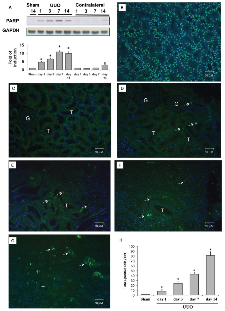
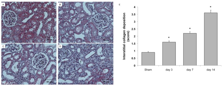
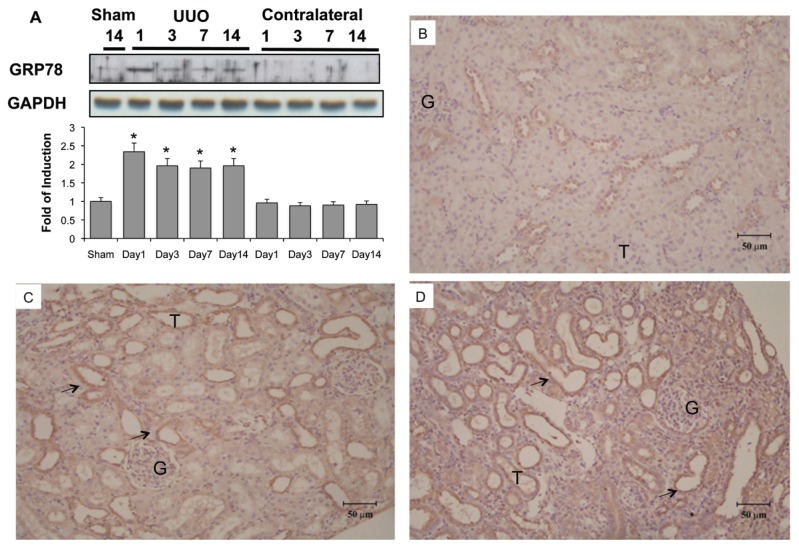
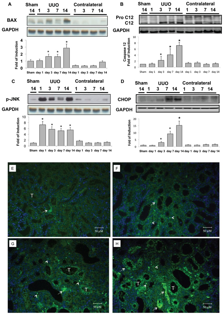
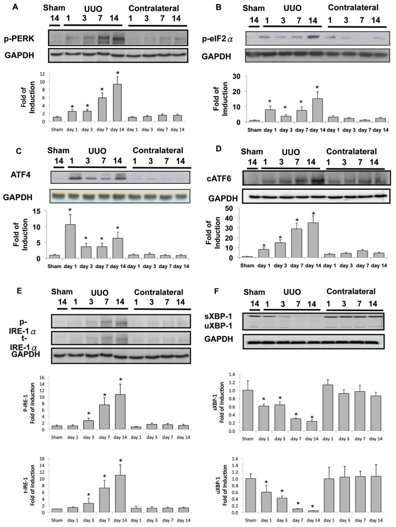
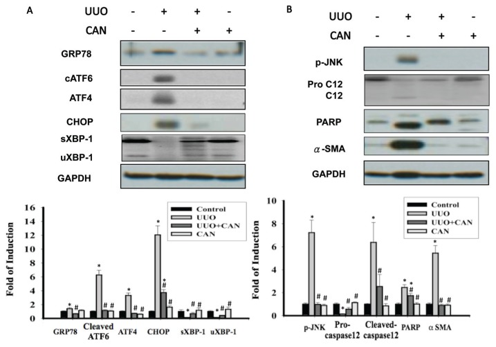
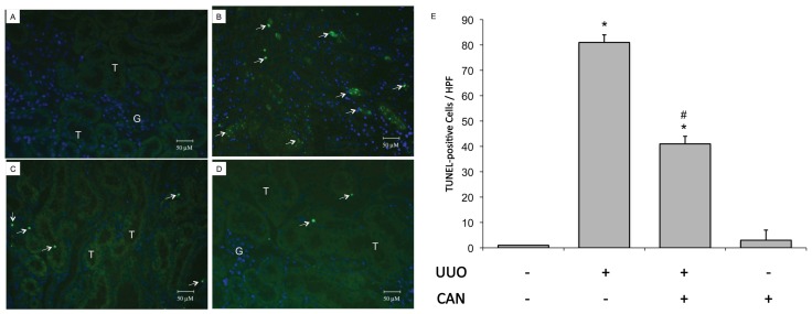
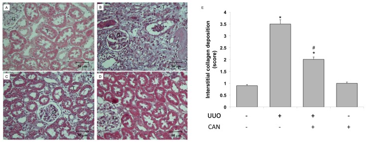
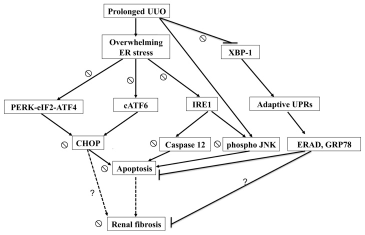
Endoplasmic reticulum (ER) stress-associated apoptosis plays a role in organ remodeling after insult. The effect of ER stress on renal tubular damage and fibrosis remains controversial. This study aims to investigate whether ER stress is involved in tubular destruction and interstitial fibrosis in vivo. Renal cell apoptosis was proven by terminal deoxynucleotidyl transferase dUTP nick end-labeling (TUNEL) stain and poly-ADP ribose polymerase expression in the unilateral ureteral obstruction (UUO) kidney. ER stress was evoked and confirmed by the upregulation of glucose-regulated protein 78 (GRP78) and the common Lys-Asp-Glu-Leu (KDEL) motif of ER retention proteins after UUO. ER stress-associated proapoptotic signals, including B-cell chronic lymphocytic leukemia (CLL)/lymphoma 2-associated × protein (BAX) expression, caspase-12 and c-Jun N-terminal kinase (JNK) phosphorylation, were activated in the UUO kidney. Prolonged ER stress attenuated both unsplicing and splicing X-box binding protein 1 (XBP-1) protein expression, but continued to activate inositol-requiring 1α (IRE1α)-JNK phosphorylation, protein kinase RNA-like endoplasmic reticulum kinase (PERK), eukaryotic translation initiation factor 2α subunit (eIF2α), activating transcription factor (ATF)-4, CCAAT/enhancer binding protein (C/EBP) homologous protein (CHOP) and cleavage activating transcription factor 6 (cATF6)-CHOP signals, which induce ER stress-related apoptosis but attenuate adaptive unfolded protein responses in UUO kidneys. However, renal apoptosis and fibrosis were attenuated in candesartan-treated UUO kidney. Candesartan was associated with maintenance of XBP-1 expression and attenuated ATF4, cATF6 and CHOP protein expression. Taken together, results show that overwhelming ER stress leads to renal cell apoptosis and subsequent fibrosis; and candesartan, at least in part, restores renal integrity by blocking ER stress-related apoptosis. Reducing ER stress may present a way to attenuate renal fibrosis.


Figures

Figure 1
Figure 1
Cleaved PARP and apoptosis in rat kidneys subjected to UUO. The left kidneys of male Wistar rats were subjected to UUO as described in Materials and Methods. Kidneys were harvested 1, 3, 7 or 14 d after UUO. (A) Cleaved PARP expression in the whole kidney lysate was evaluated via Western blot analysis using antibodies against these proteins, with GAPDH used as a loading marker. PARP was 4.7 times more upregulated 1 d after UUO and up to 10.0 times more upregulated on days 7–14. Protein bands were quantified and presented as mean ± standard error of the mean (SEM) for each group (n = 4). *P < 0.05 versus sham operation (Sham) group. Apoptosis was evaluated via a TUNEL assay. (B) DNAase-treated kidney section was used as a positive control for TUNEL staining. (C) Sham-operative kidneys show no positive nuclear stain for TUNEL. (D–G) UUO kidney showed a significant increase in TUNEL-positive staining at day 1, 3, 7 or 14. (H) The TUNEL-positive staining cells of the kidney tissue graded quantitatively as described in Materials and Methods were averaged for rats of each group (n = 4). *P < 0.05 versus the sham operation (Sham) group. The arrows indicate TUNEL-positive cells. G indicates glomerulus and T is renal tubule. 4′,6-Diamidino-2-phenylindole (DAPI) staining was used to stain the nuclei blue. HPF, high power field.
Figure 2
Figure 2
Collagen deposition in rat kidneys subjected to UUO. The left kidneys of male Wistar rats were subjected to UUO as described in Materials and Methods. At 3, 7 and 14 d after UUO, the kidneys were harvested and cut into 6-μm-thick slices for Masson trichrome staining. (A) Sham-operative kidneys showed no positive Masson trichrome staining. (B) Three days after UUO, slight tubular dilation and interstitial volume expansion was observed. Persistent ureteral obstruction induced progressive tubular dilation, interstitial volume expansion and development of fibrosis at day 7 (C) and day 14 (D) after UUO. (E) The degree of interstitial collagen deposits in Masson trichrome–stained sections of the kidney tissue graded semiquantitatively as described in Materials and Methods was averaged for rats of each group (n = 4 for each group). *P < 0.05 versus the sham operation (Sham) group.
Figure 3
Figure 3
Induction of ER chaperone and ER retention proteins in kidneys subjected to UUO. Kidneys were harvested at 1, 3, 7 or 14 d after UUO. (A) GRP78 expression in the whole kidney lysate was determined by Western blot analysis using antibodies against this protein, with GAPDH used as a loading marker. Protein bands were quantified using Image J analysis software. Values are mean ± standard error of the mean (SEM) (n = 4). *P < 0.05 versus the sham group. As compared with the sham operative kidney (B), the expression of carboxy-terminal amino acid sequence KDEL (Lys-Asp-Glu-Leu)-positive proteins were increased on the third day after UUO (C), as shown by immunohistochemical staining. KDEL-positive stained cells (arrow) were dominantly observed in the dilated tubules (D) at day 14. G indicates glomerulus and T is renal tubule.
Figure 4
Figure 4
Activation of ER stress–related proapoptotic signals in the UUO kidney. UUO gradually enhanced BAX expression (A) and increased caspase-12 in the kidney in a time-dependent manner (B). At day 14, BAX expression was 2.9 times normal, and cleavage caspase-12 was 7.2 times that of basal levels in the UUO kidney. (C) Induction of phosphorylation of JNK (p-JNK) increased by 7.3 times at 24 h after UUO. This condition lasted until the end of observation at day 14, by which time it was 5.5 times basal level. (D) UUO induced 3.0, 9.2 and 15.3 times CHOP protein expression at days 3, 7 and 14, respectively. Protein bands were quantified and normalized by GAPDH using Image J analysis software. Values are mean ± standard error of the mean (SEM) (n = 4). *P < 0.05 versus sham group. Immunofluorescence staining of CHOP was applied in the sham-operative kidneys (E) and UUO kidney at day 3 (F), day 7 (G) and day 14 (H). Progressive diffuse enhancement of CHOP fluorescence was shown in the tubular cytoplasm (arrow), and punctuated fluorescence dots localized at the nucleus (arrow head) in the UUO kidney. G indicates glomerulus and T is renal tubule.
Figure 5
Figure 5
Diminished adaptive UPR and activation of overwhelming ER stress signals in the UUO kidney. (A) PERK phosphorylation was detected at basal conditions, and UUO gradually enhanced phosphorylation to 9.3 times the basal level at day 14. eIF2α phosphorylation (B) and ATF4 protein expression (C) were induced abruptly after UUO at day 1 and continued to be expressed during the development of renal fibrosis. (D) cATF6 protein was induced by UUO. (E) Both phosphorylated and total inositol-requiring 1 (IRE1α) were enhanced after 3 d of UUO, and more significant induction of these proteins occurred at days 7 and 14 of UUO. (F) Splicing (s) and unsplicing (u) XBP-1 were both attenuated after 1 d of UUO. After 14 d of UUO, only 20% of sXBP-1 and 8% of uXBP-1 were maintained. Data are presented as mean ± standard error of the mean (SEM) (n = 3–5 per group). *P < 0.05 versus control group.
Figure 6
Figure 6
CAN reversed the activation of overwhelming ER stress signaling pathways. (A) Protein levels of GRP78, cATF6 (50 kDa), ATF4, CHOP and unsplicing and splicing XBP-1 (uXBP-1 and sXBP-1). cATF6, ATF4 and CHOP were suppressed by CAN in the UUO kidney. sXBP-1 and uXBP-1 proteins were restored by CAN in the UUO kidney. (B) Protein levels of the phosphorylated JNK, procaspase-12, PARP and α-SMA. Caspase 12, phosphorylated JNK and PARP were attenuated by CAN at day 14 in the UUO kidney. CAN also suppressed α-SMA, which suggests less renal fibrosis and epithelial-mesenchymal transition in CAN-treated kidney. Data are presented as mean ± standard error of the mean (SEM) (n = 3–5 per group). *P < 0.05 versus control group; #P < 0.05 versus UUO group.
Figure 7
Figure 7
CAN attenuated renal cell apoptosis in rat UUO kidney. Apoptosis was evaluated via a TUNEL assay. (A) Sham-operative kidneys show no positive nuclear stain for TUNEL. (B) UUO kidney showed a significant increase in TUNEL-positive staining at day 14. (C) CAN significantly attenuated UUO-induced renal cells apoptosis, as shown by TUNEL-positive staining at day 14. (D) CAN-treated sham-operative kidneys showed no significant difference compared with the sham-operative group in TUNEL staining. (E) The TUNEL-positive staining cells of the kidney tissue calculated as described in Materials and Methods were averaged for rats of each group (n = 3 for each group). G indicates glomerulus and T is renal tubule. *P < 0.05 versus the sham operation (Sham) group. #P < 0.05 versus the UUO group. The arrows indicate TUNEL-positive cells. 4′,6-Diamidino-2-phenylindole (DAPI) staining was used to stain the nuclei blue. HPF, high power field.
Figure 8
Figure 8
CAN attenuated collagen deposition in rat UUO kidney. The left kidneys of male Wistar rats were subjected to UUO as described in Materials and Methods. Oral gavage with CAN (2.5 mg/kg, twice a day) was delivered from day 1 to day 14 of UUO. At 14 d after UUO, the kidneys were harvested and cut into 6-μm-thick slices for Masson trichrome staining. (A) Sham-operative kidneys showed no positive Masson trichrome staining. Progressive tubular dilation, interstitial volume expansion and development of fibrosis were observed after 14 d of UUO (B), and CAN significantly attenuated UUO-induced interstitial damages and fibrosis (C). (D) CAN-treated sham operative did not show the interstitial damages and fibrosis. (E) The degree of interstitial collagen deposits in Masson trichrome–stained sections of the kidney tissue graded semiquantitatively as described in Materials and Methods were averaged for rats of each group (n = 3 for each group). G indicates glomerulus and T is renal tubule. *P < 0.05 versus the sham operation (Sham) group. #P < 0.05 versus the UUO group.
Figure 9
Figure 9
Schematic representation of proposed mechanisms that ER stress leading to the renal cells apoptosis and fibrosis in the UUO kidneys. Prolonged UUO activated overwhelming ER stress, including PERK-eIF2α-ATF4-CHOP, ATF6-CHOP and IRE1α-JNK/IRE1α-caspase-12 signals, leading to renal cells apoptosis. In contrast, UUO attenuated XBP-1-related adapted UPRs, which associated with antiapoptotic properties of ER-associated degradation and GRP78. Loss of renal cells is associated with inadequate tubular interstitial integrity and renal fibrosis. CAN attenuated overwhelming ER stress–related signals and restored adaptive XBP-1 activity. The dashed arrow represents the ratiocination of renal fibrosis. The ⦰ symbol indicates the action of CAN on ER stress–related signals in the UUO kidney. The question mark indicates the potential ER stress–related pathways leading to renal fibrosis in the UUO kidney.

References

    1. Hewitson TD. Renal tubulointerstitial fibrosis: common but never simple. Am J Physiol Renal Physiol. 2009;296:F1239–44. - PubMed
    1. Risdon RA, Sloper JC, De Wardener HE. Relationship between renal function and histological changes found in renal-biopsy specimens from patients with persistent glomerular nephritis. Lancet. 1968;2:363–6. - PubMed
    1. Liu Y. Renal fibrosis: new insights into the pathogenesis and therapeutics. Kidney Int. 2006;69:213–7. - PubMed
    1. Mimura I, Nangaku M. The suffocating kidney: tubulointerstitial hypoxia in end-stage renal disease. Nat Rev Nephrol. 2010;6:667–78. - PubMed
    1. Nangaku M. Chronic hypoxia and tubulointerstitial injury: a final common pathway to end-stage renal failure. J Am Soc Nephrol. 2006;17:17–25. - PubMed

Publication types

MeSH terms