. 2011;17(11-12):1275-84.
doi: 10.2119/molmed.2011.00125. Epub 2011 Aug 18.

AHR regulates WT1 genetic programming during murine nephrogenesis

Affiliations

AHR regulates WT1 genetic programming during murine nephrogenesis

M Hadi Falahatpisheh et al. Mol Med. 2011.

Abstract

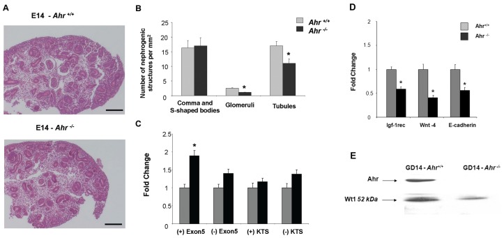
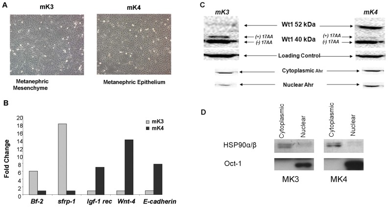
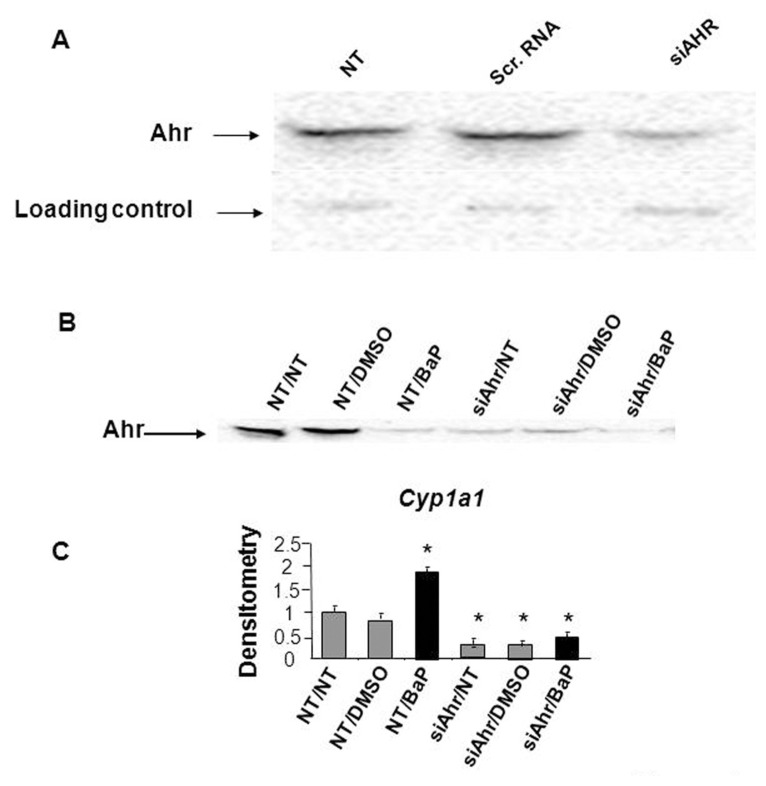
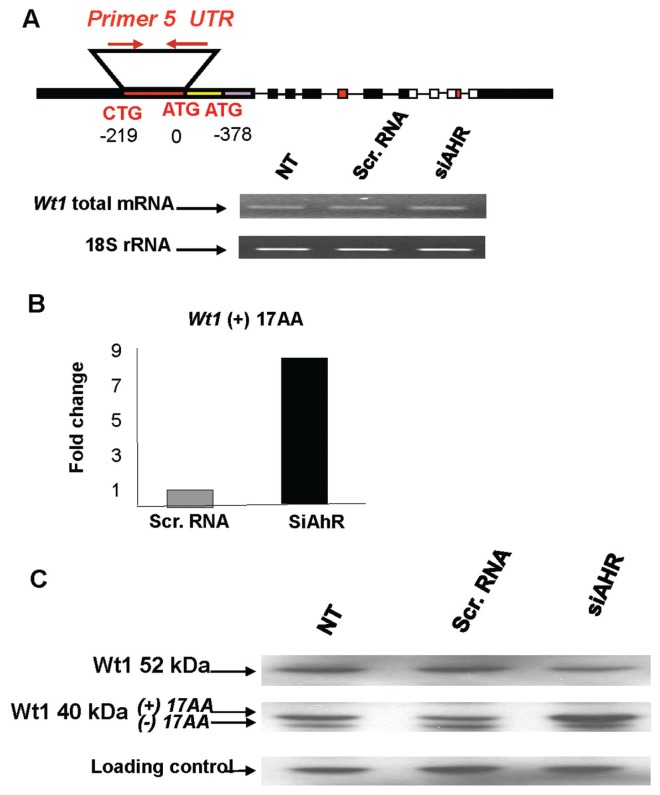
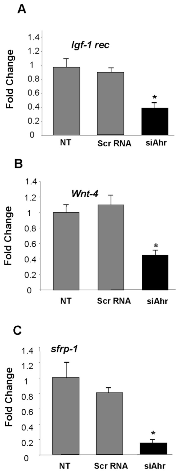
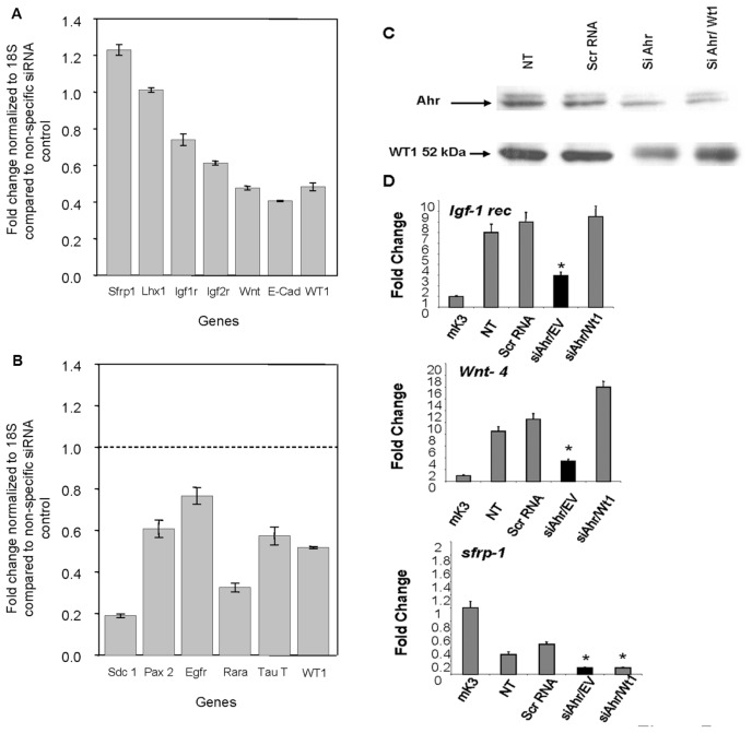
Mounting evidence suggests that the blueprint of chronic renal disease is established during early development by environmental cues that dictate alterations in differentiation programming. Here we show that aryl hydrocarbon receptor (AHR), a lig-and-activated basic helix-loop-helix-PAS homology domain transcription factor, disrupts murine renal differentiation by interfering with Wilms tumor suppressor gene (WT1) signaling in the developing kidney. Embryonic kidneys of C57BL/6J Ahr⁻/⁻ mice at gestation d (GD) 14 showed reduced condensation in the nephrogenic zone and decreased numbers of differentiated structures compared with wild-type mice. These deficits correlated with increased expression of the (+) 17aa Wt1 splice variant, decreased mRNA levels of Igf-1 rec., Wnt-4 and E-cadherin, and reduced levels of 52 kDa WT1 protein. AHR knockdown in wild-type embryonic kidney cells mimicked these alterations with notable increases in (+) 17aa Wt1 mRNA, reduced levels of 52 kDa WT1 protein, and increased (+) 17aa 40-kDa protein. AHR downregulation also reduced Igf-1 rec., Wnt-4, secreted frizzled receptor binding protein-1 (sfrbp-1) and E-cadherin mRNAs. In the case of Igf-1 rec. and Wnt-4, genetic disruption was fully reversed upon restoration of cellular Wt1 protein levels, confirming that functional interactions between AHR and Wt1 represent a likely molecular target for renal developmental interference.


Figures

Figure 1
Figure 1
Deficits in renal differentiation and Wt1 expression in embryonic kidneys from Ahr−/− mice. Kidneys were harvested from mouse embryos at GD:14 and fixed in situ for morphological examination as described. (A) Renal blastema in the proliferation zone was less condensed in developing kidneys from Ahr knockout embryos relative to wild-type counterparts (compare panels 1 and 2). Magnification in both panels was 289X. Similar findings were made in 10 mice from multiple litters. (B) Morphometric analyses of kidney sections showed decreased numbers of glomeruli and tubulo-epithelial structures (P < 0.05), despite equal numbers of comma and S-shaped bodies in Ahr knockout compared with wild-type mice. Data are presented as the average number of structures ± SD for multiple sections. Measurements were taken from serial sections from 6 to 8 embryonic kidneys isolated from three or more dams. The values shown represent the composite of serial sections with variance expressed as the difference between values for each kidney per mouse strain. (C) PCR analyses of Wt1 splice variants in embryonic kidneys of Ahr −/− compared with wild-type mice. (D) PCR analysis of various markers of renal cell differentiation (Igf-1 rec, Wnt4 and E-cadherin) in embryonic kidneys from Ahr−/− and Ahr+/+ mice. (E) Western blot analysis of Ahr and Wt1 in kidneys of Ahr−/− and Ahr+/+ mice. One representative experiment out of two is shown. Equal protein loadings were confirmed based on the signal obtained with the same antibody for a nonspecific band (80 kDa).
Figure 2
Figure 2
Ahr and Wt1 colocalize in the developing and adult kidney. Dual immunohisto-chemical analyses of Ahr and Wt1 proteins visualized using 3, 3′, 5, 5′-tetramethylbenzidine (TMB) and NovaRED, respectively (Vector Laboratories Burlingame, CA, USA). Panels A and C are representative images taken of glomeruli from adult C57BL/6J mouse kidneys or GD 11 cultured metanephros cross sections. In panel B, maximal Ahr signal (blue color) was set to threshold in serial sections processed for Ahr alone using Zeiss Axiovision Rel 4.3 image analysis. The thresholded filter was applied to dual-stained sections resulting in a red-outlined area of positive maximal Ahr signal. Ahr-Wt1 colocalization was defined by a positive nuclear Wt1 signal in proximity to the outlined maximal Ahr signal, as denoted by arrows. Because of the diffuse Ahr signal throughout metanephric glomerular cells, in panel D, Ahr-Wt1 colocalization was determined by thresholding for color resulting from both Ahr and Wt1 signals and compared with control serial sections stained for each antibody alone. The analysis showed Ahr-Wt1 colocalization outlined in red and only 10% errant signal detected in control sections. Panels E and F are serial cross sections of C57BL/6J GD 11 cultured metanephros analyzed for Ahr alone or Ahr-WT1 colocalization as described above. In panels A-D the scale represents 25 μm, while in panels E and F the scale represents 100 μm.
Figure 3
Figure 3
Differentiation and epithelialization status in mK3 and mK4 embryonic kidney cell lines. The expression of renal cell differentiation and epithelialization marker genes was examined by PCR or Western blot analysis. (A) The mK3 cells exhibited a spindle-shape morphology with irregular cytoplasmic projections, while mK4 cells displayed polygonal shapes indicative of later stage differentiation during the course of MET. (B) Real time or RT-PCR analyses showed that mK4 cells expressed higher level of E-cadherin, Wnt-4 and Igf-1 rec. compared with mK3 cells. In contrast Bf-2 and secreted frizzled receptor binding protein-1 (sfrp-1) transcripts were higher in mK3 cells. Similar results were seen in three different experiments. (C) Western blot analysis showed that mK4 cells possess higher levels of Ahr and 52 kDa Wt1 protein compared with mK3 cells. Similar results were seen in four separate experiments. (D) The quality of cellular fractions was confirmed by Western blotting using HSP90α/β and Oct1 as markers of cytoplasmic and nuclear proteins, respectively. Images shown are representative of triplicate samples from multiple gels. Equal protein loadings were confirmed based on the signal obtained with the same antibody for a nonspecific band (80 kDa). Notably, embryonic kidney cell lines also expressed 40 kDa variants of ± exon 5. The abundance of (+) 17 AA variant inversely correlated with the degree of renal cell differentiation as shown by decreases in Bf-2 and sfrp-1 mRNAs and increases in Igf-1 rec, Wnt-4 and E-cadherin mRNAs.
Figure 4
Figure 4
Downregulation of Ahr by siRNA in the mK4 embryonic kidney cell line. (A) Ahr protein levels decreased in mK4 cells transfected with siRNA directed at the AhR, while protein levels remain unchanged in cells treated with scrambled (scr) RNA as a nonspecific negative siRNA control. NT = no treatment. One representative experiment from a total of ten is shown. Equal protein loadings were confirmed based on the signal obtained with the same antibody for a nonspecific band (25 kDa). (B) Hydrocarbon inducibility of Cyp1a1 is abolished in mK4 cells following Ahr siRNA transfection. (C) RT-PCR for Cyp1a1 shows that only siRNA against Ahr inhibits Cyp1a1 inducibility by BaP. One representative experiment out of three is shown. Densitometry is shown in arbitrary units. * denotes statistical differences from respective NT controls.
Figure 5
Figure 5
Regulation of Wt1 by Ahr. Expression profiles of Wt1 mRNA or protein were examined in mK4 cells following siRNA Ahr knockdown. (A) RT-PCR was performed using a primer set that hybridizes to the Wt1 5′ UTR region upstream of the translation start sites for the 52 and 40 kDa isoforms. Ahr knockdown did not alter total Wt1 mRNA levels. The signal for 18S rRNA is shown as a normalization control. (B) Real time PCR was performed using primer sets specific for the (±) KTS and (±) 17 AA Wt1 splice variants. Scr = scrambled. AhR knockdown caused a pronounced increase in (+) 17 AA Wt1 expressions in mK4 cells. (C) Western blot analysis showed that Ahr knockdown caused selective decreases in 52 kDa Wt1 protein isoform and corresponding increases in 40 kDa isoforms. A nonspecific ~80-kDa protein recognized by the Wt1 antibody was used as a loading control. Similar results were seen in four different experiments.
Figure 6
Figure 6
Ahr plays a regulatory role in programming of MET and differentiation of mK4 cells. Real time PCR was performed using specific primers for various molecular targets to determine the expression of effector genes essential in nephrogenesis. Ahr knockdown in mK4 caused a significant decreases in mRNA levels for Igf-1 rec. (A), Wnt-4 (B), and sfrp-1 (C) (* P < 0.05). One representative experiment out of three is shown. NT = no treatment; Scr = scrambled.
Figure 7
Figure 7
Interactions between Ahr and Wt1 during the course of renal cell differentiation. (A) Downregulation of markers of differentiation by Wt1 siRNA. mK4 cells were cultured in the presence of Wt1 siRNA for 3 d. qRT-PCR values were calculated to represent fold change normalized to 18S compared with nonspecific siRNA control. sfrp-1 and lim 1 homeobox gene were used as markers of mesenchymal identity, while Igf-1 rec, Igf-2 rec, Wnt4, and E-cadherin were used as markers of epithelial identity. (B) Deregulation of Wt1 targets by Wt1 siRNA. mK4 cells were cultured in the presence of Wt1 siRNA for 3 d. qRT-PCR values were calculated to represent fold change normalized to 18S compared with nonspecific siRNA control. Syndecan1, paired box protein 2, EGF rec, retinoic acid receptor alpha, taurine transporter, and Wilms tumor transcription factor. (C) The mK4 cells were transfected for 24 h with the CB6-Wt1 (−) exon 5/(−) exon 9 plasmid encoding for 52 kDa Wt1 protein or empty vector after transfection with Ahr siRNA. Reduced Wt1 protein levels following Ahr knockdown were rescued in cells transfected with Wt1 cDNA compared with empty vector. (D) Real time PCR shows decreased mRNA levels for Igf-1 rec. and Wnt-4 following Ahr knockdown and effective rescue in cells transfected with Wt1 cDNA; however, decreases in sFrp-1 mRNA following Ahr knockdown were not restored in cells transfected with Wt1 cDNA. One representative experiment out of three is shown. NT = no treatment.

Similar articles

Cited by

References

    1. Barker DJ, Winter PD, Osmond C, Margetts B, Simmonds SJ. Weight in infancy and death from ischaemic heart disease. Lancet. 1989;2:577–80. - PubMed
    1. Lim K, Armitage JA, Stefanidis A, Oldfield BJ, Black MJ. Intrauterine growth restriction in the absence of postnatal ‘catch-up’ growth leads to improved whole body insulin sensitivity in rat offspring. Pediatr Res 2011 - PubMed
    1. Menendez-Castro C, et al. Early and late postnatal myocardial and vascular changes in a protein restriction rat model of intrauterine growth restriction. PLoS One. 2011;6:e20369. - PMC - PubMed
    1. Nanez A, Ramos KS. Introduction and Overview of Receptor Systems. In: McQueen CA, editor. Comprehensive Toxicology. 2nd ed. Vol. 2. Elsevier; San Diego: 2010. pp. 71–80. editor in chief. Available at: http://www.knovel.com/web/portal/browse/display?_EXT_KNOVEL_DISPLAY_book....
    1. Falahatpisheh MH, Ramos KS. Ligand-activated Ahr signaling leads to disruption of nephrogenesis and altered Wilms’ tumor suppressor mRNA splicing. Oncogene. 2003;22:2160–71. - PubMed

Publication types

MeSH terms