. 2011 Oct 4;50(39):8323-32.
doi: 10.1021/bi2009322. Epub 2011 Sep 6.

Crystallographic trapping of heme loss intermediates during the nitrite-induced degradation of human hemoglobin

Affiliations

Crystallographic trapping of heme loss intermediates during the nitrite-induced degradation of human hemoglobin

Jun Yi et al. Biochemistry. .

Abstract

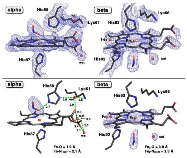
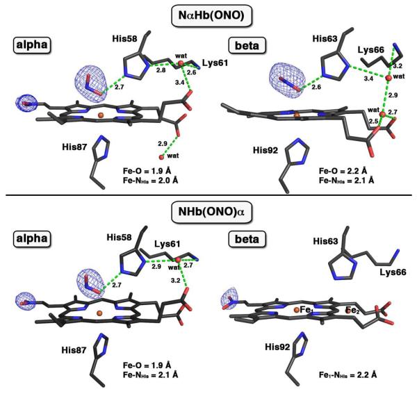
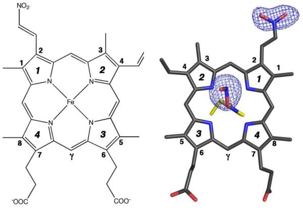
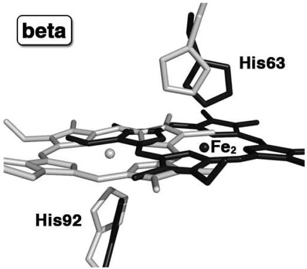
Heme is an important cofactor in a large number of essential proteins and is often involved in small molecule binding and activation. Loss of heme from proteins thus negatively affects the function of these proteins but is also an important component of iron recycling. The characterization of intermediates that form during the loss of heme from proteins has been problematic, in a large part, because of the instability of such intermediates. We have characterized, by X-ray crystallography, three compounds that form during the nitrite-induced degradation of human α(2)β(2) hemoglobin (Hb). The first is an unprecedented complex that exhibits a large β heme displacement of 4.8 Å toward the protein exterior; the heme displacement is stabilized by the binding of the distal His residue to the heme Fe, which in turn allows for the unusual binding of an exogenous ligand on the proximal face of the heme. We have also structurally characterized complexes that display regiospecific nitration of the heme at the 2-vinyl position; we show that heme nitration is not a prerequisite for heme loss. Our results provide structural insight into a possible pathway for nitrite-induced loss of heme from human Hb.


Figures

Figure 1
Figure 1
(Top panel) The 2Fo-Fc electron density maps (contoured at 1.0σ) and final models of the α- and β-heme active sites of the ferric human Hb(ONO)d,p structure (PDB accession code: 3ONZ, 2.1 Å resol.). (Bottom panel) The Fo-Fc omit electron density map (bottom, contoured at 3.0σ) and final models of the α- and β-heme active sites of this compound. The H-bonding interactions are shown in green dashed lines. The Fo-Fc omit electron density maps for water molecules in the α subunit are not shown here. Bonds to Fe are omitted for clarity.
Figure 2
Figure 2
Fo-Fc omit electron density maps (contoured at 3.0σ) and final model of the α- and β-heme active sites of two ferric human NHb nitrite adducts. The top panel is for NαHb(ONO) (PDB accession code: 3OO4, 1.9 Å resol.), and the bottom panel is for NHb(ONO)α (PDB accession code: 3OO5, 2.1 Å resol.). The two Fe atoms in the β subunit of NHb(ONO)α structure were modeled with an Fe–Fe distance of 4.6 Å. The H-bonding interactions are shown in green dashed lines, and bonds to Fe are omitted for clarity.
Figure 3
Figure 3
(Left) The commonly used Fisher’s numbering system for the protoporphyrin IX macrocycle. (Right) Top view of the Fo-Fc omit electron density map (contoured at 3.0σ) and final model of the a nitriheme and Fe-ONO moieties of NαHb(ONO). The proximal His87 residue is shown in yellow.
Figure 4
Figure 4
The β heme sites of the superposed final models of our previously published HbONO structure (PDB accession code: 3D7O, 1.8 Å resol., shown in light gray) and new Hb(ONO)d,p structure (PDB accession code: 3ONZ, 2.1 Å resol., shown in black); for clarity, the Fe1 and nitrite ligands are not shown.
Figure 5
Figure 5
The heme vinyl regions of the α subunit of ferric aquometHb (PDB accession code 3P5Q, 2.0 Å resol.). The close contacts between the terminal Cβ atoms at two vinyl positions of the α heme and the protein backbone Cα atoms of the nearby residues (top panel) and the side chains of nearby residues (bottom panel) are shown as yellow dashed lines. (A) The close contacts of the terminal Cβ atom at the 4-vinyl position with the Cα atoms of nearby helices (e.g Helix C and Helix G); (B) The close contacts of the terminal Cβ atom at the 2-vinyl position with the Cα atoms of nearby helices (Helix E, Helix G and Helix H); (C) The close contacts of the terminal Cβ atom at the 4-vinyl position with the side chains of nearby residues; (D) The close contacts of the terminal Cβ atom at the 2-vinyl position with the side chains of nearby residues.

Similar articles

Cited by

References

    1. Yonetani T, Laberge M. Protein Dynamics Explain the Allosteric Behaviors of Hemoglobin. Biochim. Biophys. Acta. 2008;1784:1146–1158. - PMC - PubMed
    1. Boon EM, Marletta MA. Ligand Specificity of H-NOX Domains: From sGC to Bacterial NO Sensors. J. Inorg. Biochem. 2005;99:892–902. - PubMed
    1. Guengerich FP, editor. Human Cytochrome P450 Enzymes. 3rd Ed Kluwer Academic/Plenum; New York: 2005.
    1. Hargrove MS, Singleton EW, Quillin ML, Ortiz LA, Phillips GN, Olson JS, Mathews AJ. His(64)(E7)→Tyr Apomyoglobin as a Reagent for Measuring Rates of Hemin Dissociation. J. Biol. Chem. 1994;269:4207–4214. - PubMed
    1. Hargrove MS, Wilkinson AJ, Olson JS. Structural Factors Governing Hemin Dissociation from Metmyoglobin. Biochemistry. 1996;35:11300–11309. - PubMed

Publication types