. 2011 Aug 24:8:105.
doi: 10.1186/1742-2094-8-105.

Air pollution & the brain: Subchronic diesel exhaust exposure causes neuroinflammation and elevates early markers of neurodegenerative disease

Affiliations

Air pollution & the brain: Subchronic diesel exhaust exposure causes neuroinflammation and elevates early markers of neurodegenerative disease

Shannon Levesque et al. J Neuroinflammation. .

Abstract

Background: Increasing evidence links diverse forms of air pollution to neuroinflammation and neuropathology in both human and animal models, but the effects of long-term exposures are poorly understood.

Objective: We explored the central nervous system consequences of subchronic exposure to diesel exhaust (DE) and addressed the minimum levels necessary to elicit neuroinflammation and markers of early neuropathology.

Methods: Male Fischer 344 rats were exposed to DE (992, 311, 100, 35 and 0 μg PM/m³) by inhalation over 6 months.

Results: DE exposure resulted in elevated levels of TNFα at high concentrations in all regions tested, with the exception of the cerebellum. The midbrain region was the most sensitive, where exposures as low as 100 μg PM/m³ significantly increased brain TNFα levels. However, this sensitivity to DE was not conferred to all markers of neuroinflammation, as the midbrain showed no increase in IL-6 expression at any concentration tested, an increase in IL-1β at only high concentrations, and a decrease in MIP-1α expression, supporting that compensatory mechanisms may occur with subchronic exposure. Aβ42 levels were the highest in the frontal lobe of mice exposed to 992 μg PM/m³ and tau [pS199] levels were elevated at the higher DE concentrations (992 and 311 μg PM/m³) in both the temporal lobe and frontal lobe, indicating that proteins linked to preclinical Alzheimer's disease were affected. α Synuclein levels were elevated in the midbrain in response to the 992 μg PM/m³ exposure, supporting that air pollution may be associated with early Parkinson's disease-like pathology.

Conclusions: Together, the data support that the midbrain may be more sensitive to the neuroinflammatory effects of subchronic air pollution exposure. However, the DE-induced elevation of proteins associated with neurodegenerative diseases was limited to only the higher exposures, suggesting that air pollution-induced neuroinflammation may precede preclinical markers of neurodegenerative disease in the midbrain.


Figures
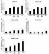
Figure 1
Figure 1
Subchronic DE Exposure Elevates TNFα in the Brain: Midbrain Vulnerability. Male Fischer 344 rats were exposed to either filtered air (control, 0 μg PM/m3 DE, n = 8), 35 μg PM/m3 DE (Low, n = 8), 100 μg PM/m3 DE (Mid Low, n = 8), 311 μg PM/m3 DE (Mid High, n = 8), or 992 μg PM/m3 DE (High, n = 8) for 6 months. TNFα protein levels from the (A) Midbrain, (B) Olfactory Bulbs, (C) Temporal Lobe, (D) Frontal Lobe, and (E) Cerebellum were measured by ELISA. An * indicates significant difference (p < 0.05) from control animals. While all components of the brain, with the exception of the cerebellum, showed an elevated TNFα response to DE at some concentration of DE, the midbrain was the most sensitive, producing a significant increase from control at only 100 μg PM/m3. = DE.
Figure 2
Figure 2
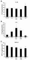
Subchronic DE Exposure Differentially Regulates Other Cytokines and Chemokines in the Midbrain. Male Fischer 344 rats were exposed to either filtered air (control, 0 μg PM/m3 DE, n = 8), 35 μg PM/m3 DE (Low, n = 8), 100 μg PM/m3 DE (Mid Low, n = 8), 311 μg PM/m3 DE (Mid High, n = 8), or 992 μg PM/m3 DE (High, n = 8) for 6 months. (A) IL-1β, (B) IL-6, and (C) MIP-1α protein levels were measured in the midbrain by ELISA. An * indicates significant difference (p < 0.05) from control animals. DE elevated IL-1β at only the highest concentration of DE, failed to affect IL-6 levels, and decreased MIP-1α expression in the midbrain.
Figure 3
Figure 3
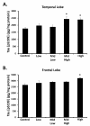
Subchronic DE Exposure Elevates Tau [pS199] in the Temporal and Frontal Lobes. Male Fischer 344 rats were exposed to either filtered air (control, 0 μg PM/m3 DE, n = 8), 35 μg PM/m3 DE (Low, n = 8), 100 μg PM/m3 DE (Mid Low, n = 8), 311 μg PM/m3 DE (Mid High, n = 8), or 992 μg PM/m3 DE (High, n = 8) for 6 months. Tau [pS199] protein levels were measured in the (A) Frontal and (B) Temporal lobe by ELISA. An * indicates significant difference (p < 0.05) from control animals. DE elevated Tau [pS199] at the highest concentrations of DE, demonstrating that subchronic exposure to high levels of air pollution is associated with Alzheimer disease-like pathology.
Figure 4
Figure 4
Subchronic DE Exposure Elevates α Synuclein in the Midbrain. Male Fischer 344 rats were exposed to either filtered air (control, 0 μg PM/m3 DE, n = 8), 35 μg PM/m3 DE (Low, n = 8), 100 μg PM/m3 DE (Mid Low, n = 8), 311 μg PM/m3 DE (Mid High, n = 8), or 992 μg PM/m3 DE (High, n = 8) for 6 months. α Synuclein protein levels were measured in the midbrain by western blot. An * indicates significant difference (p < 0.05) from control animals. DE elevated α synuclein protein levels in the midbrain at only the highest concentrations tested, demonstrating that subchronic exposure to high levels of air pollution is associated with Parkinson's disease-like pathology.
Figure 5
Figure 5
Subchronic DE Exposure Elevates Aβ in the Frontal Lobe. Male Fischer 344 rats were exposed to either filtered air (control, 0 μg PM/m3 DE, n = 8), 35 μg PM/m3 DE (Low, n = 8), 100 μg PM/m3 DE (Mid Low, n = 8), 311 μg PM/m3 DE (Mid High, n = 8), or 992 μg PM/m3 DE (High, n = 8) for 6 months. Aβ42 protein levels were measured in the frontal lobe ELISA. An * indicates significant difference (p < 0.05) from control animals. DE elevated Aβ42 protein levels in the frontal lobe at only the highest concentrations tested, demonstrating that subchronic exposure to high levels of air pollution is associated with Alzheimer's disease-like pathology.

Similar articles

Cited by

References

    1. Block ML, Zecca L, Hong JS. Microglia-mediated neurotoxicity: uncovering the molecular mechanisms. Nat Rev Neurosci. 2007;8:57–69. doi: 10.1038/nrn2038. - DOI - PubMed
    1. Glass CK, Saijo K, Winner B, Marchetto MC, Gage FH. Mechanisms underlying inflammation in neurodegeneration. Cell. 2010;140:918–934. doi: 10.1016/j.cell.2010.02.016. - DOI - PMC - PubMed
    1. Frank-Cannon TC, Alto LT, McAlpine FE, Tansey MG. Does neuroinflammation fan the flame in neurodegenerative diseases? Mol Neurodegener. 2009;4:47. doi: 10.1186/1750-1326-4-47. - DOI - PMC - PubMed
    1. Carvey PM, Punati A, Newman MB. Progressive dopamine neuron loss in Parkinson's disease: the multiple hit hypothesis. Cell Transplant. 2006;15:239–250. doi: 10.3727/000000006783981990. - DOI - PubMed
    1. Horowitz MP, Greenamyre JT. Gene-environment interactions in Parkinson's disease: the importance of animal modeling. Clin Pharmacol Ther. 2010;88:467–474. doi: 10.1038/clpt.2010.138. - DOI - PMC - PubMed

Publication types

MeSH terms