

Marrow cell genetic phenotype change induced by human lung cancer cells
- PMID: 21864488
- PMCID: PMC4568832
- DOI: 10.1016/j.exphem.2011.08.008
Marrow cell genetic phenotype change induced by human lung cancer cells
Abstract
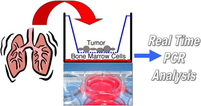
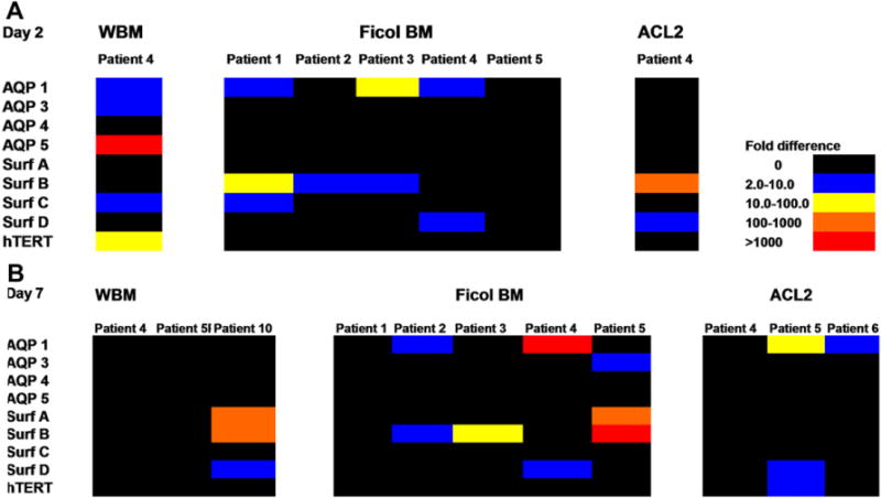
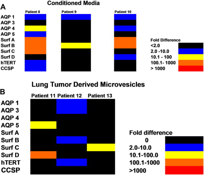
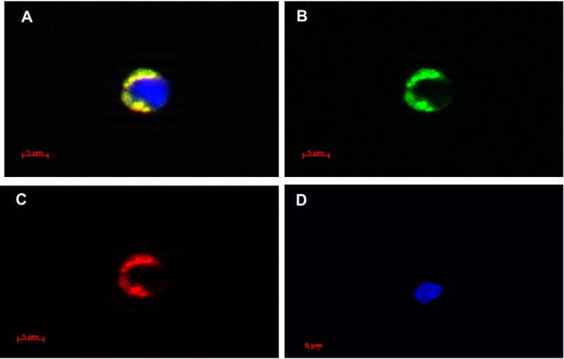
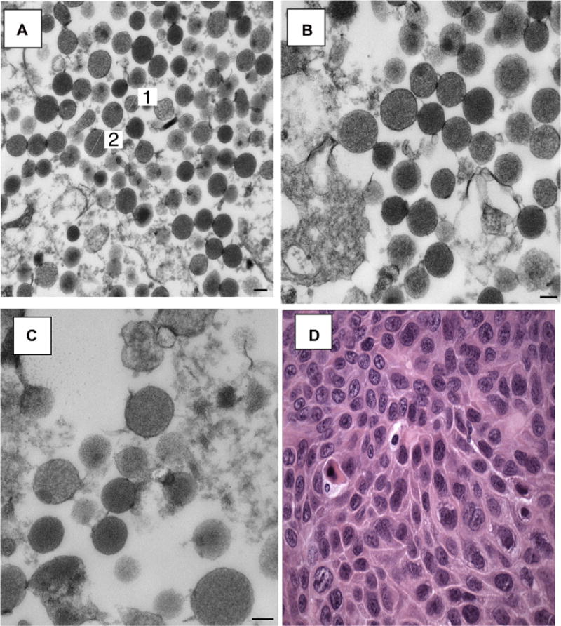
Microvesicles have been shown to mediate varieties of intercellular communication. Work in murine species has shown that lung-derived microvesicles can deliver mRNA, transcription factors, and microRNA to marrow cells and alter their phenotype. The present studies evaluated the capacity of excised human lung cancer cells to change the genetic phenotype of human marrow cells. We present the first studies on microvesicle production by excised cancers from human lung and the capacity of these microvesicles to alter the genetic phenotype of normal human marrow cells. We studied 12 cancers involving the lung and assessed nine lung-specific mRNA species (aquaporin, surfactant families, and clara cell-specific protein) in marrow cells exposed to tissue in co-culture, cultured in conditioned media, or exposed to isolated lung cancer-derived microvesicles. We assessed two or seven days of co-culture and marrow which was unseparated, separated by ficoll density gradient centrifugation or ammonium chloride lysis. Under these varying conditions, each cancer derived from lung mediated marrow expression of between one and seven lung-specific genes. Microvesicles were identified in the pellet of ultracentrifuged conditioned media and shown to enter marrow cells and induce lung-specific mRNA expression in marrow. A lung melanoma and a sarcoma also induced lung-specific mRNA in marrow cells. These data indicate that lung cancer cells may alter the genetic phenotype of normal cells and suggest that such perturbations might play a role in tumor progression, tumor recurrence, or metastases. They also suggest that the tissue environment may alter cancer cell gene expression.
Copyright © 2011. Published by Elsevier Inc.
Figures
References
-
- Morel O, Toti F, Hugel B, Freyssinet JM. Cellular microparticles: a disseminated storage pool of bioactive vascular effectors. Curr Opin Hematol. 2004;11:156–164. - PubMed
-
- Janowska-Wieczorek A, Majka M, Kijowski J, et al. Platelet-derived microparticles bind to hematopoietic stem/progenitor cells and enhance their engraftment. Blood. 2001;98:3143–3149. - PubMed
-
- Baj-Krzyworzeka M, Majka M, Pratico D, et al. Platelet-derived microparticles stimulate proliferation, survival, adhesion, and chemotaxis of hematopoietic cells. Exp Hematol. 2002;30:450–459. - PubMed
-
- Rozmyslowicz T. Platelet- and megakaryocyte-derived microparticles transfer CXCR4 receptor to CXCR4-null cells and make them susceptible to infection by X4-HIV. AIDS. 2003;17:33–42. - PubMed
-
- Graves LE, Ariztia EV, Navari JR, Matzel HJ, Stack MS, Fishman DA. Proinvasive properties of ovarian cancer ascites-derived membrane vesicles. Cancer Res. 2004;64:7045–7049. - PubMed
Publication types
MeSH terms
Substances
Grants and funding
LinkOut - more resources
Full Text Sources
Medical
