Review
. 2012 Jan;83(1):3-11.
doi: 10.1016/j.mvr.2011.08.001. Epub 2011 Aug 16.

Cross talk between focal adhesion kinase and cadherins: role in regulating endothelial barrier function

Affiliations
Review

Cross talk between focal adhesion kinase and cadherins: role in regulating endothelial barrier function

Sadiqa K Quadri. Microvasc Res. 2012 Jan.

Abstract

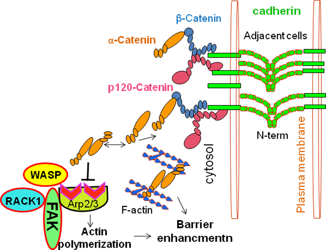
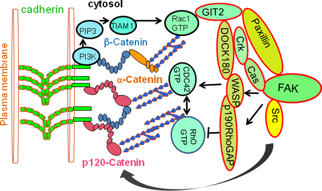
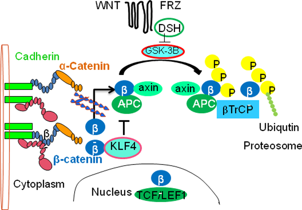
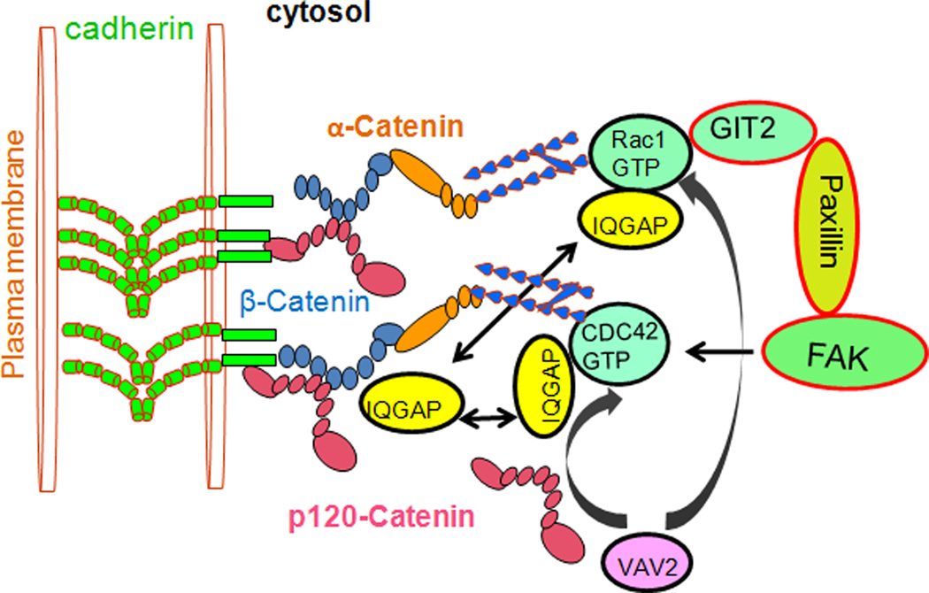
A layer of endothelial cells attached to their underlying matrices by complex transmembrane structures termed focal adhesion (FA) proteins maintains the barrier property of microvascular endothelium. FAs sense the physical properties of the extracellular matrix (ECM) and organize the cytoskeleton accordingly. The close association of adherens junction (AJ) protein, cadherin, with the cytoskeleton is known to be essential in coordinating the appropriate mechanical properties to cell-cell contacts. Recently, it has become clear that a crosstalk exists between focal adhesion kinase (FAK) and cadherin that regulates signaling at intercellular endothelial junctions. This review discusses recent advances in our understanding of the dynamic regulation of the molecular connections between FAK and the cadherin complex and cadherin-catenin-actin interaction-dependent changes as well as the role of small GTPases in endothelial barrier regulation. This review also discusses how a signaling network regulates a range of cellular processes important for barrier function and diseases.


Figures

Fig. 1
Fig. 1. Model of FAK and the cadherin complex function in actin polymerization
FAK signals to Arp2/3 in modulating cell adhesion and actin polymerization. The FAK N-terminal domain binds directly to Arp3, which promotes nucleation by bringing an actin monomer to Arp2/3 via the WASP-homology 2 domain, thereby enhancing actin polymerization. The α-catenin isoform binds to actin preferentially as a dimer, and the α-catenin monomer binds to β-catenin; therefore α-catenin does not bind to β-catenin and actin simultaneously.
Fig. 2
Fig. 2. Structure of strand-swapping in wild-type E-cadherin fragments (trans-dimer)
Ribbon diagram shows the strand-swapped dimer formed between protomers of wild-type E-cadherin EC1 in the crystal. Side chain atoms are shown for Trp2 residues (W2) and calcium ions are displayed as green spheres (reproduced with permission of the Nature Structure Molecular Biology Harrison, 2010).
Fig. 3
Fig. 3. Interactions between FAK and the cadherin complex
Signal from FAK to Rac1 plays a role in modulating cell adhesion and actin polymerization. PI3K is recruited to the membrane by intact E-cadherin AJs, where it generates PIP3, resulting in the activation of the Rho-GEF, TIAM1 and subsequently of Rac1 and CDC42. Activated FAK enhances Rac1 activity via a Cas/CrkII/DOCK180 complex. Paxillin binds to GIT2, participate in Rac- and Rho-mediated signaling events at FAs. Association of β-catenin with paxillin is depends on Rac and Cdc42 activities. Src family Activates p190 Rho-GAP, activated p190 Rho-GAP suppresses Rho activity. p120 is a Src substrate and is involved in the maintenance of cadherin at the plasma membrane.
Fig. 4
Fig. 4. Signaling pathways affected by loss of cadherin function
Upon disassembly of the cytoplasmic cell-adhesion complex, catenins are released and accumulate in the cytoplasm. β-Catenin (β) is then sequestered by the adenomatous polyposis coli (APC)–axin–glycogen synthase kinase 3β (GSK-3β) complex and phosphorylated by GSK-3β. Phosphorylated β-catenin is specifically bound by βTrCP, a subunit of the E3 ubiquitin-ligase complex, which ubiquitylates β-catenin and thereby marks it for rapid proteosomal degradation. However, on activation of the WNT signalling pathway, GSK-3β is repressed, and β-catenin is no longer phosphorylated. β-catenin translocates to the nucleus where, together with the TCF/LEF1 transcription factors, it modulates the expression of several target genes. Transcriton factor KLF4 interacts with β-catenin and inhibits Wnt signaling pathway.
Fig 5
Fig 5. Cross talk between FAK and cadherin
Since β-catenin does not contain FAK or paxillin binding sites, FAK signals to Rac, activated Rac1 and CDC42 sequester the GTPase-activating protein IQGAP1, and prevent its binding to β-catenin, thereby stabilizing cadherin-mediated cell adhesion. Otherwise in free form IQGAP1 binds to β-catenin, thereby displacing α-catenin from the cytoplasmic adhesion complex and disrupting the anchoring of the cytoplasmic complex to the cytoskeleton. Cytoplasmic p120-catenin (p120) activates the Rho-family GTPases Rac1 and CDC42 through the VAV2 (Rho-GEF), and represses Rho by an unknown mechanism.

Similar articles

Cited by

References

    1. Abercrombie M, Dunn GA. Adhesions of fibroblasts to substratum during contact inhibition observed by interference reflection microscopy. Exp Cell Res. 1975;92:57–62. - PubMed
    1. Abercrombie M, et al. The locomotion of fibroblasts in culture. IV. Electron microscopy of the leading lamella. Exp Cell Res. 1971;67:359–367. - PubMed
    1. Aberle H, et al. Assembly of the cadherin-catenin complex in vitro with recombinant proteins. J Cell Sci. 1994;107(Pt 12):3655–3663. - PubMed
    1. Al-Amoudi A, Frangakis AS. Structural studies on desmosomes. Biochem Soc Trans. 2008;36:181–187. - PubMed
    1. Anastasiadis PZ, Reynolds AB. The p120 catenin family: complex roles in adhesion, signaling and cancer. J Cell Sci. 2000;113(Pt 8):1319–1334. - PubMed

Publication types

MeSH terms