. 2012 May 15:61:132-43.
doi: 10.1016/j.visres.2011.08.007. Epub 2011 Aug 16.

Neural bases of selective attention in action video game players

Affiliations

Neural bases of selective attention in action video game players

D Bavelier et al. Vision Res. .

Abstract

Over the past few years, the very act of playing action video games has been shown to enhance several different aspects of visual selective attention, yet little is known about the neural mechanisms that mediate such attentional benefits. A review of the aspects of attention enhanced in action game players suggests there are changes in the mechanisms that control attention allocation and its efficiency (Hubert-Wallander, Green, & Bavelier, 2010). The present study used brain imaging to test this hypothesis by comparing attentional network recruitment and distractor processing in action gamers versus non-gamers as attentional demands increased. Moving distractors were found to elicit lesser activation of the visual motion-sensitive area (MT/MST) in gamers as compared to non-gamers, suggestive of a better early filtering of irrelevant information in gamers. As expected, a fronto-parietal network of areas showed greater recruitment as attentional demands increased in non-gamers. In contrast, gamers barely engaged this network as attentional demands increased. This reduced activity in the fronto-parietal network that is hypothesized to control the flexible allocation of top-down attention is compatible with the proposal that action game players may allocate attentional resources more automatically, possibly allowing more efficient early filtering of irrelevant information.


Figures

Figure 1
Figure 1
Example of stimuli. Subjects had to indicate with a button press which of two targets (square or diamond) appeared in the ring of shapes while maintaining fixation on the centre cross. In the low load condition the remaining seven shapes in the annulus were circles and in the high load condition the remaining shapes were a mixture of circles and other shapes (e.g., triangles, trapezoids, houses). The distractors, patches of moving or static dots, were placed on both the left and right, either inside (central distractors) or outside (peripheral distractors) the annulus of shapes.
Figure 2
Figure 2
Sample scanning run. Each scanning run lasted 5 minutes and included eight, 36-second blocks as well as an initial dummy block of 12 seconds (not depicted in the figure). In each run there were four blocks of the two load levels presented in a randomized block order. During each block the distractors (patches of dots) were alternately moving or static in 18-second intervals. The moving or static state of the dots was also randomized within a block. There were eight scanning runs: 4 with central distractors and 4 with peripheral distractors. Run order was randomized across subjects.
Figure 3
Figure 3
Behavioral results. RT data plotted by load level with % correct data noted on each bar for the peripheral distractor and the central distractor conditions respectively. There was no significant difference across load level or distractor eccentricity in terms of accuracy (percentages at the base of the histograms), but there was a main effect of load on reaction time. In both experiments, the high load condition produced longer RTs. Importantly, VGPs and NVGPs were comparably slowed down from low to high load, indicating similar increase in difficulty across groups.
Figure 4
Figure 4
Pattern of activation as attentional load is increased for NVGPs (a) and for VGPs (b). VGPs show much reduced recruitment of the fronto-parietal network as compared to NVGPs (see p. 11 for statistical parameters).
Figure 5
Figure 5
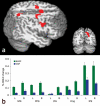
Greater activation for NVGPs than VGPs was noted as load increased in areas of the fronto-parietal network (a). Difference in percent bold changes between high and low load is plotted for five regions of interest in the attentional network system in NVGPs and in VGPs (b). SFS = Superior Frontal Sulcus, MFG = Middle Frontal Gyrus, IFG = Inferior Frontal Gyrus, Cing = Cingulum, IPS = Intraparietal Sulcus.
Figure 6
Figure 6
Pattern of activation for central versus peripheral distractors computed through a conjunction analysis of VGPs and NVGPs activation maps (see p. 12, top paragraph for statistical parameters). Across groups, greater activation for central distractors was noted more posteriorly along the calcarine sulcus, whereas greater activation for peripheral distractors was observed more anteriorly, as predicted by the known retinotopic organization of early visual areas.
Figure 7
Figure 7
(a) Percent BOLD signal change in area MT/MST as a function of load and the eccentricity of the irrelevant motion patch. (b) MT/MST ROI for one representative subject.

Similar articles

Cited by

References

    1. Bavelier D, Tomann A, Hutton C, Mitchell T, Liu G, Corina D, Neville H. Visual attention to periphery is enhanced in congenitally deaf individuals. Journal of neuroscience. 2000;20:1–6. - PMC - PubMed
    1. Beauchamp M, Cox R, DeYoe E. Graded effects of spatial and featural attention on human area MT and associated motion processing areas. Journal of Neurophysiology. 1997;78(1):516–520. - PubMed
    1. Beauchamp MH, Dagher A, Aston JAD, Doyon J. Dynamic functional changes associated with cognitive skill learning of an adapted version of the Tower of London task. NeuroImage. 2003;20(3):1649–1660. - PubMed
    1. Beck DM, Lavie N. Look here but ignore what you see: effects of distractors at fixation. Journal of Experimental Psychology: Human Perception and Performance. 2005;31(3):592–607. - PubMed
    1. Beckmann C, Jenkinson M, Smith SM. General multi-level linear modelling for group analysis in FMRI. NeuroImage. 2003;20:1052–1063. - PubMed

Publication types