. 2011 Dec;25(8):2010-6.
doi: 10.1016/j.tiv.2011.08.001. Epub 2011 Aug 16.

Mitochondrial permeability and toxicity of diethylhexyl and monoethylhexyl phthalates on TK6 human lymphoblasts cells

Affiliations

Mitochondrial permeability and toxicity of diethylhexyl and monoethylhexyl phthalates on TK6 human lymphoblasts cells

Carlos A Rosado-Berrios et al. Toxicol In Vitro. 2011 Dec.

Abstract

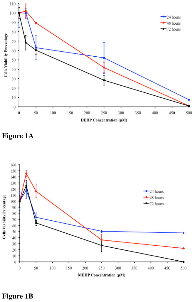
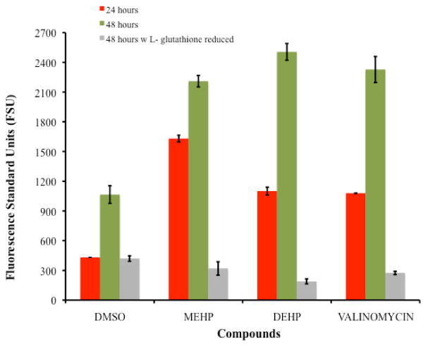
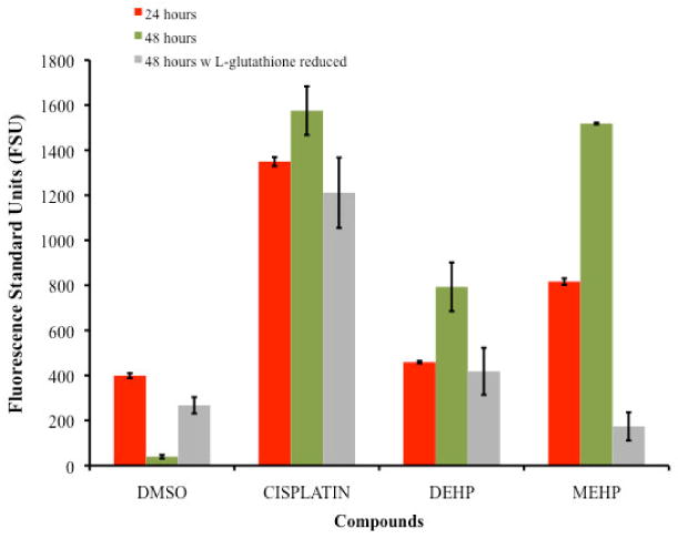
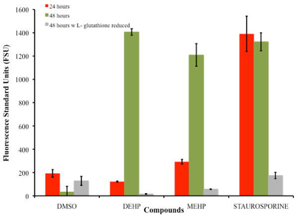
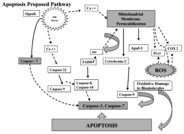
Phthalates are ubiquitous compounds used in the manufacturing industry. Some are known endocrine disruptors, acting as xenoestrogens, others induce reproductive toxicity and damage to DNA among other effects. Studies on apoptosis induction and mitochondrial damage capacity of phthalates on the immune system are limited. This study aims to determine cell viability inhibition and apoptosis induction of diethylhexyl phthalate (DEHP) and monoethylhexyl phthalate (MEHP) on the human TK6 lymphoblast cell line at concentrations found in the environment. Key hallmark events, such as mitochondrial membrane permeability, generation of reactive oxygen species (ROS) and activation of caspase 3 and 7 were measured. Concentrations that inhibit viability of 50% (IC50) of the cells were determined at 24, 48 and 72 h with doses ranging from 10 to 500 μM. Changes in mitochondrial membrane permeability, ROS generation and activation of caspases 3 and 7, were measured as part of the cell death mechanism. The IC50 at 24 h was approximately 250 μM for both phthalates; at 48 h were 234 and 196 μM for DEHP and MEHP, respectively and at 72 h IC50s were 100 and 80 μM for DEHP and MEHP, respectively. Overall the longer the time of exposure the lower the IC50's for both compounds. Both compounds affected mitochondrial membrane potential, promoted ROS generation and activated caspases 3 and 7. MEHP is more toxic, promotes higher level of ROS production and caspases activation. Our findings suggest that DEHP and MEHP have the capacity to induce apoptosis in cells of the immune system at concentrations found in the environment.


Conflict of interest statement

Conflict of Interests: The authors declare they have no actual or potential competing financial interests.

Figures

Figure 1
Figure 1
Figure 1A. TK6 cells viability after 24, 48 and 72 hours of exposure to DEHP. The 24 hours IC50 was 250μM, the 48 hours IC50 was 234μM and the 72 hours IC50 was 100μM. Figure 1B. TK6 cells viability after 24, 48 and 72 hours exposure to MEHP. The 24 hours IC50 was 250μM, the 48 hours IC50 was 196μM and the 72 hours IC50 was 80μM.
Figure 2
Figure 2
Comparison of changes in the mitochondrial membrane potential among TK6 cells exposed for 24 and 48 hours to the IC50's of DEHP (234μM) and MEHP (196μM). For the 48 hours exposure assay the average level of mitochondrial membrane permeability caused by DEHP and MEHP presented P-values of 0.002 and 0.024, respectively when compared with the negative control. Also no significant difference was observed among the positive control (valinomycin) with MEHP and DEHP (P-value >0.10). Comparison of the changes in the mitochondrial membrane potential generated on TK6 cells exposed for 48 hours to the IC50's of DEHP (234μM) and MEHP (196μM) with 500μM of L- glutathione is also presented an illustrates the reduction effect.
Figure 3
Figure 3
Comparison of the Generation of Reactive Oxygen Species as detected by DCFH-DA of TK6 cells exposed for 24 and 48 hours to the IC50's of DEHP (234μM) and MEHP (196μM). For the 48 hours exposure assay there is significant difference (p<0.05) in the level of ROS generated by the cells exposed to DEHP compared with the positive control and MEHP. The cells exposed to DEHP and MEHP also showed significant differences (P-value = 0.009). But no significant difference was observed among the positive control (cisplatin) and MEHP (P-value >0.1). Comparison of the ROS generated of TK6 cells exposed for 48 hours to the IC50's of DEHP (234μM) and MEHP (196μM) with 500μM of L- glutathione reduced are also presented also presented an illustrates the reduction in ROS generation.
Figure 4
Figure 4
Comparison of Caspases 3 and 7 Activation of TK6 cells exposed for 48 hours to the IC50's of DEHP (234μM) and MEHP (196μM). For the 48 hours exposure period the comparison of the positive control staurosporine with DEHP produced a significance value of 1.0, and with MEHP a value of 0.999 indicating no significant difference in the level of caspases 3 and 7 activation between DEHP, MEHP and the positive control. Comparison of Caspases 3 and 7 Activation on TK6 cells exposed for 48 hours to the IC50's of DEHP (234μM) and MEHP (196μM) with 500μM of L- glutathione reduced are also presented an illustrates the reduction effect.
Figure 5
Figure 5
Proposed cell death mechanism induced on TK6 cells after treatment with DEHP and MEHP. Events presented in this pathway are a summary of general events involved in apoptosis. As demonstrated in this study the cell death mechanism induced by DEHP and MEHP includes mitochondrial membrane permeability, generation of ROS and activation of caspases 3 and 7 (shaded shapes). Segmented lines indicate apoptotic contributing events that were not measured in this study but are proposed for DEHP and MEHP based on our findings. The differences observed in mitochondrial permeability, ROS and caspase activation levels generated by MEHP and DEHP imply that different factors are influencing their cell death mechanism. Although not tested in this study phthalates have shown to stimulate Ca2+ entry in the cells and mitochondria which increases ROS production as well as significant crossover between the two principal apoptosis pathways suggesting the existence of positive feedback loops (Palleschi et al. 2009; Bissonnette et al. 2008). The difference between the ROS generated by MEHP and DEHP may be caused also by the release and/or activation of specific mitochondrial proteins such as Prx3, COX-2 and cytochrome c oxidase.

Similar articles

Cited by

References

    1. Beauchesne I, Barnabe S, Cooper DG, Nicell JA. Plasticizers and related toxic degradation products in wastewater sludges. Water Science & Technology. 2008;57(3):367–374. doi: 10.2166/wst.2008.001. - DOI - PubMed
    1. Bissonnette SL, Teague JE, Sherr DH, Schlezinger JJ. An endogenous prostaglandin enhances environmental phthalate-induced apoptosis in bone marrow B cells: Activation of distinct but overlapping pathways. Journal of Immunology. 2008;181:1728–1736. - PMC - PubMed
    1. Bornehag CG, Sundell J, Weschler CJ, Sigsgaard T, Lundgren B, Hasselgren M, Hägerhed-Engman L. The association between asthma and allergic symptoms in children and phthalates in house dust: A nested case-control study. Environmental Health Perspective. 2004;112:1393–1397. doi: 10.1289/ehp.7187. - DOI - PMC - PubMed
    1. Brunelle JK, Letai A. Control of mitochondrial apoptosis by the Bcl-2 family. Journal of Cell Science. 2009;122:437–441. doi: 10.1242/jcs.031682. - DOI - PMC - PubMed
    1. Carter JE, Roll DB, Petersen RV. The in Vitro hydrolysis of di-(2- ethylhexyl) phthalate by rat tissues. Drug Metabolism and Disposition. 1974;2(4):341–344. - PubMed

Publication types

MeSH terms