. 2012 Apr 7;9(69):657-64.
doi: 10.1098/rsif.2011.0370. Epub 2011 Aug 24.

Direct evidence of phospholipids in gecko footprints and spatula-substrate contact interface detected using surface-sensitive spectroscopy

Affiliations

Direct evidence of phospholipids in gecko footprints and spatula-substrate contact interface detected using surface-sensitive spectroscopy

Ping Yuan Hsu et al. J R Soc Interface. .

Abstract

Observers ranging from Aristotle to young children have long marvelled at the ability of geckos to cling to walls and ceilings. Detailed studies have revealed that geckos are 'sticky' without the use of glue or suction devices. Instead, a gecko's stickiness derives from van der Waals interactions between proteinaceous hairs called setae and substrate. Here, we present surprising evidence that although geckos do not use glue, a residue is transferred on surfaces as they walk-geckos leave footprints. Using matrix-free nano-assisted laser desorption-ionization mass spectrometry, we identified the residue as phospholipids with phosphocholine head groups. Moreover, interface-sensitive sum-frequency generation spectroscopy revealed predominantly hydrophobic methyl and methylene groups and the complete absence of water at the contact interface between a gecko toe pad and the substrate. The presence of lipids has never been considered in current models of gecko adhesion. Our analysis of gecko footprints and the toe pad-substrate interface has significant consequences for models of gecko adhesion and by extension, the design of synthetic mimics.


Figures

Figure 1.
Figure 1.
The invisible and hydrophobic residue of a gecko's step left behind on the substrate. (a) Picture of a gecko (Gekko vittatus) sticking on a glass substrate. (b) After the gecko walked on the clean glass, the glass panel was placed in a room at 98% humidity, allowing moisture to condense on the glass surface. The glass panel became opaque as a result of tiny water droplets covering the surface. The outline of footprints was revealed by differential condensation around the footprints. The picture was collected using a digital camera. Similar footprints were also observed for Gekko gecko and Anolis carolinensis (electronic supplementary material, figure S10). (Online version in colour.)
Figure 2.
Figure 2.
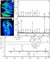
Mass spectroscopy analysis and imaging of the gecko footprint. (a) The NALDI mapping image of the peak m/z 184 collected after the gecko toe was brought in contact with the NALDI plate immediately after shedding so that the new setae were exposed. The increased brightness (intensity) in the seta-bearing scansors corresponds to higher concentrations of the substance yielding the m/z 184 signal. (b) The scan of the ions observed at m/z 600–1000 in the NALDI spectrum. The spatial distributions of several ions in the NALDI spectrum were mapped and they also show similar images to that obtained for m/z 184 (see electronic supplementary material, figures S2 and S7). For results shown in (c,d), the geckos were housed for three weeks after their moulting cycle in a glass cage, then their feet were cleaned by touching them many times on a clean glass substrate. The NALDI mass spectrum was collected after this process by touching the gecko toe on the NALDI substrate. (c) Mapping of the m/z 184 ion signal, whereas (d) ions observed in the range from m/z 600 to 1000. (e) Tandem mass spectrum of m/z 734. The major fragment peak at m/z 184 corresponds to phosphocholine head group. The remaining neutral chain (without the head group) appears as a weak peak at m/z 551. Further, the m/z 478 and 496 peaks indicate the loss of one of C16 fatty acids tails from precursor and its dehydrated form, respectively. The fragmentation pattern identifies m/z 734 as phosphatidylcholine 16 : 0/16 : 0.
Figure 3.
Figure 3.
Probing the molecular structure of the contact interface between the gecko toe and the sapphire substrate. (a) The SFG spectra collected after bringing the gecko toe in contact with a sapphire prism. (b) The sapphire surface was wetted with water and the SFG spectrum was collected after bringing the gecko toe in contact with the sapphire substrate. (c) The gecko toe was peeled off the dry substrate and the SFG spectrum was collected of the residue left behind on the sapphire substrate. (d) The SFG spectrum of a clean sapphire surface (without any exposure to water) shows no discernible peak in the hydrocarbon and water region. All the SFG spectra are collected using SSP polarization (s-polarized SFG, s-polarized infrared and p-polarized visible beam). Solid lines are fit to a Lorentzian equation described elsewhere [15]. (e) Schematic of the SFG experimental measurement. The infrared beam (black line) and the visible beam (red line) were overlapped at the contact interface between toe and sapphire, the SFG beam (orange line) was then analysed. The SFG peaks in the range from 2800 to 3000 cm-1 correspond to stretching vibrations of methyl and methylene groups, and the range from 3200 to 3800 cm-1 correspond to stretching vibrations of water.
Figure 4.
Figure 4.
Models visualizing the location of lipid molecules on the spatula. In the homogeneous model (a), lipid material (yellow) may form an evenly distributed layer encasing the spatular structure. This would be equivalent to a lipid membrane covering keratin fibres (red). However, our results cannot exclude a heterogeneous model for contact (b) and (c), where the lipid material is not responsible for all the contact at the interface, allowing either lipid material or keratin protein to make contact with the solid substrate. Within the heterogeneous model there are two possibilities. In (b) the lipid molecules form the inter-layer between the two keratin rods or in (c) the lipid material is evenly distributed with the keratin proteins.

References

    1. Autumn K., Liang Y. A., Hsieh S. T., Zesch W., Chan W. P., Kenny T. W., Fearing R., Full R. J. 2000. Adhesive force of a single gecko foot-hair. Nature 405, 681–68510.1038/35015073 (doi:10.1038/35015073) - DOI - DOI - PubMed
    1. Autumn K., et al. 2002. Evidence for van der Waals adhesion in gecko setae. Proc. Natl Acad. Sci. USA 99, 12 252–12 25610.1073/pnas.192252799 (doi:10.1073/pnas.192252799) - DOI - DOI - PMC - PubMed
    1. Irschick D. J., Herrel A., Vanhooydonck B. 2006. Whole-organism studies of adhesion in pad-bearing lizards: creative evolutionary solutions to functional problems. J. Comp. Physiol. A Neuroethol. Sens. Neural Behav. Physiol. 192, 1169–117710.1007/s00359-006-0145-2 (doi:10.1007/s00359-006-0145-2) - DOI - DOI - PubMed
    1. Arzt E., Gorb S., Spolenak R. 2003. From micro to nano contacts in biological attachment devices. Proc. Natl Acad. Sci. USA 100, 10 603–10 60610.1073/pnas.1534701100 (doi:10.1073/pnas.1534701100) - DOI - DOI - PMC - PubMed
    1. Geim A. K., Dubonos S. V., Grigorieva I. V., Novoselov K. S., Zhukov A. A., Shapoval S. Y. 2003. Microfabricated adhesive mimicking gecko foot-hair. Nat. Mater. 2, 461–46310.1038/nmat917 (doi:10.1038/nmat917) - DOI - DOI - PubMed

Publication types

LinkOut - more resources