. 2012 Apr 7;9(69):640-7.
doi: 10.1098/rsif.2011.0395. Epub 2011 Aug 24.

Tilted cellulose arrangement as a novel mechanism for hygroscopic coiling in the stork's bill awn

Affiliations

Tilted cellulose arrangement as a novel mechanism for hygroscopic coiling in the stork's bill awn

Yael Abraham et al. J R Soc Interface. .

Abstract

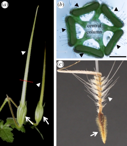
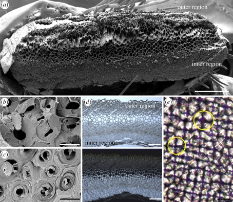
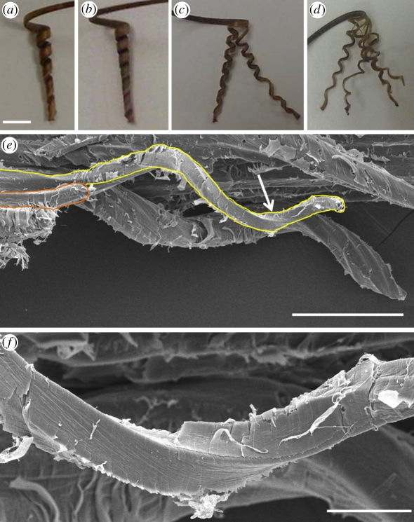
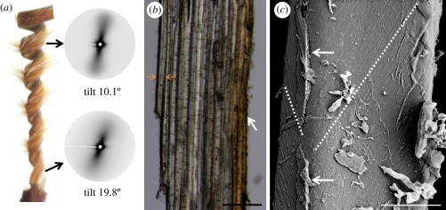
The sessile nature of plants demands the development of seed-dispersal mechanisms to establish new growing loci. Dispersal strategies of many species involve drying of the dispersal unit, which induces directed contraction and movement based on changing environmental humidity. The majority of researched hygroscopic dispersal mechanisms are based on a bilayered structure. Here, we investigate the motility of the stork's bill (Erodium) seeds that relies on the tightening and loosening of a helical awn to propel itself across the surface into a safe germination place. We show that this movement is based on a specialized single layer consisting of a mechanically uniform tissue. A cell wall structure with cellulose microfibrils arranged in an unusually tilted helix causes each cell to spiral. These cells generate a macroscopic coil by spiralling collectively. A simple model made from a thread embedded in an isotropic foam matrix shows that this cellulose arrangement is indeed sufficient to induce the spiralling of the cells.


Figures

Figure 1.
Figure 1.
The morphology of the fruit of stork's bill (Erodium gruinum). (a) Two complete stork's bill-shaped fruits, about 4 days prior to ripening. Arrows indicate the location of the seed; arrowheads indicate the awns. Dashed red line indicates the part from which cross section (b) was taken. (b) Erodium gruinum fruit in cross section depicts five awns (indicated by arrowheads) connected by a central column. (c) Dry awned seed showing the coiling region (arrowhead) close to the seed (arrow). Scale bar, (b) 1 mm.
Figure 2.
Figure 2.
Microscopic images of the awn cross section at the coiling region. (a) Overview of a fractured cross section taken by scanning electron microscope. (b) A close up of the coil outer layer reveals a brittle break morphology. (c) A close up of the inner layer reveals spool-like packing of the cellulose fibrils, which is typical of the whole layer. (d) Light (upper panel) and polarized light (lower panel) microscopy images of a 10 µm thick cross section from the coiling region of the awn. Under crossed polarizers, the part facing the inner side of the coil is brighter, indicating a relatively high microfibril angle. (e) A close up of the cells in the inner layer, revealing a dark cross which is typical of circular birefringent materials (such as starch granules [25]). Yellow circles delineate the margins of a cell. Scale bars, (a) 250 µm, (b,c,e) 10 µm and (d) 100 µm.
Figure 3.
Figure 3.
Cooperative cell spiralling creates the macroscopic coil. (a) The coiling section of the complete awn showing five to six coils. (b) The separated inner layer of the awn, showing seven to eight coils. The inner layer, split into (c) once and (d) twice still coils to about the same extent as the complete inner layer (the distortions of some of the sections result from the unevenness of the cuts). (e) Scanning electron micrograph of the inner layer of the awn showing a group of coiling cells behind a single coiled cell connected to the tissue at one end (delineated). (f) A close up of the cell region is indicated by an arrow in (e). Scale bars, (ad) 5 mm, (e) 100 µm and (f) 20 µm.
Figure 4.
Figure 4.
The cellulose microfibrils organization in the cell walls of the coiling cells. (a) Small-angle X-ray scattering (SAXS) pattern of a vertical sample from the inner layer of the stork's bill awn, measured at the top and bottom parts of the coiling region. The tighter coil in the bottom part shows a larger SAXS tilt. (b) Longitudinal section of the inner layer showing the cells' alignment with the length of the awn. As the length of single cells is about 1 mm, it is impossible to see complete cells in this view. (c) Cryo-scanning electron image showing the change in microfibril angle in a single cell, marked by a broken line. Arrows indicate remains of the middle lamella. Scale bars, (b) 100 µm, and (c) 5 µm.
Figure 5.
Figure 5.
Schematic showing the arrangement of cellulose microfibril in a normal helix (a) common in elongated plant cell, compared with a tilted helical arrangement of the cellulose microfibrils in the coiling cells of the stork's bill awn (b). In the common plant cell, the cell axis (in red) coincides with the cellulose helix, so that the microfibril angle (MFA) between the cellulose and the cell axis does not change with the circumference of the cell. On the other hand, in the spiralling cells of the stork's bill, the helix axis (in yellow) is at an angle to the cell axis (in red), resulting in the changing of the MFA around the cell. (c) A scheme illustrating the identical cell polarity in the inner layer of the awn at the coiling region. The direction of the tilting of the cellulose helix is the same in the cells, with the largest microfibril angle facing towards the wide side of the awn.
Figure 6.
Figure 6.
Sponge models simulating the difference in behaviour of drying plant cell walls with normal and tilted helix alignment of the cellulose microfibrils. Long rectangular cylinder sponge strips (approx. 1.5 × 1.5 × 12 cm3) were threaded loosely to form a helix, either tilted (b–d) or not (f). The thread was then tightened, forming a non-extendable cage restricting the expansion of the isotropic matrix of the sponge. (a) A scheme (right: three-dimensional, left-sides projections) illustrating the threading angles of the tilted helix models: β on side II, and δ on side IV, whereas sides I and III have α = γ= 0°. In (b) tight anticlockwise thread helix with angles of β = +15°, δ = −8° and (c) β = +40°, δ = −33.5° results in a clockwise coiling of the structure. Note that the coiling radius of the structure is reduced in model (c) with the increase in tilt angles. (d) For a clockwise thread helix of β = −40°, and δ = +33.5°, an anticlockwise coil is formed. (e) A scheme (right: three-dimensional, left-sides projections) illustrating the threading angles in the normal helix models. All the sides are threaded at the same angle, α. (f) A normal thread helix with α = +10° produces only a twist with no bending of the structure's longitudinal axis. Scale bar, (be,f) 2 cm.

References

    1. Fowler N. L. 1988. What is a safe site? Neighbor, litter, germination date, and patch effects. Ecology 69, 947–961 10.2307/1941250 (doi:10.2307/1941250) - DOI
    1. Van der Pijl L. 1982. Principles of dispersal in higher plants, 3rd edn. New York, NY: Springer
    1. Fahn A., Werker E. 1972. Anatomical mechanisms of seed dispersal. In Seed biology (ed. Kozlowski T. T.), pp. 151–221 New York, NY: Academic Press
    1. Harper J. L., Lovell P. H., Moore K. G. 1970. The shapes and sizes of seeds. Annu. Rev. Ecol. Syst. 1, 327–356 10.1146/annurev.es.01.110170.001551 (doi:10.1146/annurev.es.01.110170.001551) - DOI
    1. Elbaum R., Zaltzman L., Burgert I., Fratzl P. 2007. The role of wheat awns in the seed dispersal unit. Science 316, 884–886 10.1126/science.1140097 (doi:10.1126/science.1140097) - DOI - PubMed

Publication types