. 2011 Oct 20;118(16):4377-83.
doi: 10.1182/blood-2011-02-338053. Epub 2011 Aug 24.

Impaired FcεRI stability, signaling, and effector functions in murine mast cells lacking glycosylphosphatidylinositol-anchored proteins

Affiliations

Impaired FcεRI stability, signaling, and effector functions in murine mast cells lacking glycosylphosphatidylinositol-anchored proteins

Wouter L W Hazenbos et al. Blood. .

Abstract

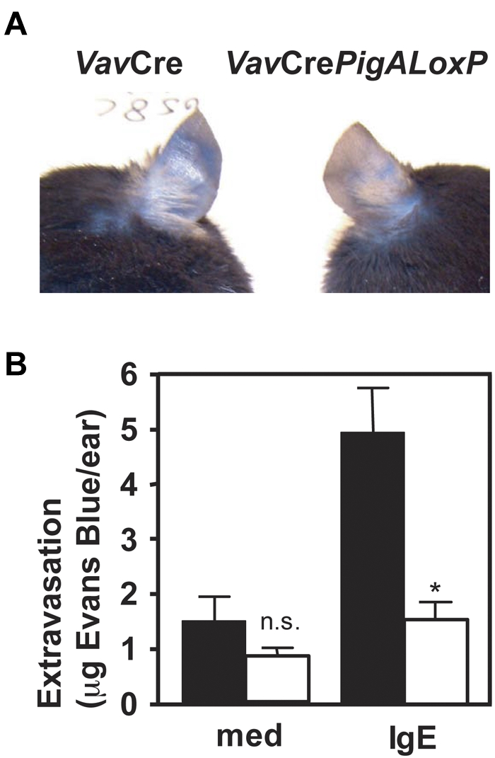
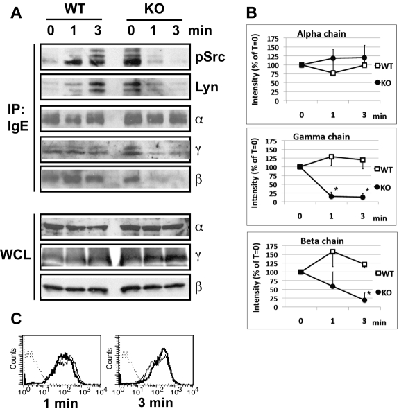
A key event and potential therapeutic target in allergic and asthmatic diseases is signaling by the IgE receptor FcεRI, which depends on its interactions with Src family kinases (SFK). Here we tested the hypothesis that glycosylphosphatidylinositiol-anchored proteins (GPI-AP) are involved in FcεRI signaling, based on previous observations that GPI-AP colocalize with and mediate activation of SFK. We generated mice with a hematopoietic cell-specific GPI-AP deficiency by targeted disruption of the GPI biosynthesis gene PigA. In these mice, IgE-mediated passive cutaneous anaphylaxis was largely abolished. PigA-deficient mast cells cultured from these mice showed impaired degranulation in response to stimulation with IgE and antigen in vitro, despite normal IgE binding and antigen-induced FcεRI aggregation. On stimulation of these cells with IgE and antigen, coprecipitation of the FcεRI α-chain with the γ-chain and β-chain was markedly reduced. As a result, IgE/antigen-induced FcεRI-Lyn association and γ-chain tyrosine phosphorylation were both impaired in PigA-deficient cells. These data provide genetic evidence for an unanticipated key role of GPI-AP in FcεRI interchain interactions and early FcεRI signaling events, necessary for antigen-induced mast cell degranulation.


Figures

Figure 1
Figure 1
Phenotype analysis of PigA-deficient mast cells by flow cytometry. (A) PigA-deficient BMMCs cultured from VavCrePigALoxP (KO; bold lines) or littermate control VavCre (WT; filled lines) mice were analyzed by flow cytometry. Complete GPI-deficient phenotype was evident from a lack of staining with proaerolysin, a general marker for the GPI lipid anchor, and with antibodies against the GPI-AP CD48 and HSA. PigA-deficient BMMCs demonstrated normal staining with antibodies against the mast cell marker c-Kit, and with the lipid raft marker cholera toxin subunit B (CTx); and normal binding of monomeric mouse IgE. Dotted lines: staining controls. (B) Peritoneal mast cells from littermate control VavCre or VavCrePigALoxP mice were stained with anti–c-Kit and proaerolysin; c-Kit positive cells were gated in forward/side scatter. Negative proaerolysin staining demonstrates in vivo mast cell GPI-deficiency; this was confirmed using antibodies against CD48 and HSA (not shown).
Figure 2
Figure 2
Resistance to IgE-mediated passive cutaneous anaphylaxis in mice with PigA-deficient mast cells. Mice were given injections with IgE anti-DNP in one ear and as control with medium in the other. On the next day, intravenous injections of DNP-BSA mixed with Evans Blue were given. Extravasation was visualized by blue staining of the ears (top panel). After extraction from the ears, extravasated Evans blue was quantified (bottom panel). Filled bars: VavCre littermate control mice; open bars: VavCrePigALoxP mice. Results represent mean ± SEM of 3 mice per group. Asterisk indicates significant difference (P < .01) with control mice; n.s. indicates not significant.
Figure 3
Figure 3
Impaired IgE/antigen-mediated degranulation of PigA-deficient BMMCs. BMMCs were (A) sensitized with IgE anti-DNP, followed by challenge with various concentrations of DNP-BSA for 1 hour, or (B) activated with the nonspecific degranulation stimulus compound 48/80. Degranulation of the cells was expressed as the percentage specific release of β-hexosaminidase into the supernatant relative to the total cell content. In the absence of IgE sensitization, the mean specific β-hexosaminidase release was below 3% for both control and PigA-deficient BMMC at either concentration of DNP-BSA tested (not shown). Filled bars: littermate control BMMC; open bars: PigA-deficient BMMC. Results represent mean ± SEM of at least 3 experiments; asterisks indicate significant difference (P < .05) with control cells.
Figure 4
Figure 4
Normal IgE/antigen-induced FcϵRI aggregation in PigA-deficient BMMCs. BMMCs were sensitized with IgE anti-DNP, and either left in medium (-Ag) or challenged with DNP-BSA antigen (+Ag) for 3 minutes, fixed and stained with fluorescent anti–mouse IgG(H+L). (A) Confocal microscope images show the appearance of FcϵRI aggregates at the upper cell surface after antigen challenge (+Ag) in both control (WT; top panels) and PigA-deficient BMMCs (KO; bottom panels). (B) Quantification of the number of FcϵRI aggregates per cell by the Particle Analysis software. Shown are the mean numbers ± SEM particles per cell, as a measure for FcϵRI aggregation, of at least 15 cells per group. Filled bars: littermate control BMMC; open bars: PigA-deficient BMMCs. Asterisk indicates significant difference (P < .05) with control cells; n.s. indicates not significant.
Figure 5
Figure 5
Reduced IgE/antigen induced FcϵRI-SFK assoctiation and FcϵRI interchain interactions in PigA-deficient BMMCs. (A) Littermate control (WT) or PigA-deficient (KO) BMMCs were sensitized with IgE anti-DNP for 3 hours at 37°C, and either mixed with DNP-BSA on ice (0 minutes), or further incubated with prewarmed DNP-BSA at 37°C for 1 or 3 minutes before transfer to lysis buffer on ice. Top panels: FcϵRI receptor complexes were immunoprecipitated from lysates by anti-IgE (IP: IgE), and immunoblotted with antibodies against FcϵRI α-chain (α), γ-chain (γ), or β-chain (β); or against Lyn (Lyn) or activated SFK (pSrc). In the pSrc blot, the two bottom bands presumably represent Lyn, the upper band Fyn, based on their molecular weight values (53/56 kDa and 59 kDa, respectively). Bottom panels: whole cell lysates (WCL) were immunoblotted with antibodies against FcϵRI α-chain (α), γ-chain (γ), or β-chain (β); data representative of at least two experiments are shown. (B) Quantitative scanning of multiple coimmunoprecipitation experiments specified in panel A, confirming significantly reduced coprecipitation of the γ-chain and β-chain with the receptor complex on stimulation. Shown are signal intensities of anti-IgE immunoprecipitates which were immunoblotted with antibodies against FcϵRI γ-chain (middle panel; n = 4) or β-chain (bottom panel; n = 3); or against α-chain as control (top panel; n = 3). Data are expressed as mean percentages of pixel intensities ± SEM, compared with nonstimulated cells (T = 0). Asterisks indicate significant differences (P < .05) compared with WT cells. (C) IgE binding to control (filled lines) or PigA-deficient (bold lines) BMMCs after IgE sensitization followed by 1 or 3 minutes antigen stimulation.
Figure 6
Figure 6
Reduced IgE/antigen-induced FcR γ-chain tyrosine phosphorylation in PigA-deficient BMMCs. After sensitization of BMMCs with IgE anti-DNP for 3 hours at 37°C, DNP-BSA (Ag) was added and the cells were challenged for 3 minutes at 37°C, or left on ice (0 minutes), and lysed. Top panel: immunoprecipitates prepared using anti–γ-chain antibodies were immunoblotted with anti-phosphotyrosine antibody 4G10 (γ-pY), or as loading control with anti–γ-chain (γ). Bottom panel: lysates were immunoprecipitated using anti-phosphotyrosine antibody 4G10 and immunoblotted with anti–β-chain antibodies (β-pY); loading controls are lysates immunoblotted with anti–β-chain antibodies (β). WT: littermate control BMMC; KO: PigA-deficient BMMCs; data representative of at least 2 experiments are shown.

Similar articles

Cited by

References

    1. Turner H, Kinet JP. Signalling through the high-affinity IgE receptor Fc epsilonRI. Nature. 1999;402(6760 Suppl):B24–30. - PubMed
    1. Gilfillan AM, Rivera J. The tyrosine kinase network regulating mast cell activation. Immunol Rev. 2009;228(1):149–169. - PMC - PubMed
    1. Field KA, Holowka D, Baird B. Compartmentalized activation of the high affinity immunoglobulin E receptor within membrane domains. J Biol Chem. 1997;272(7):4276–4280. - PubMed
    1. Stauffer TP, Meyer T. Compartmentalized IgE receptor-mediated signal transduction in living cells. J Cell Biol. 1997;139(6):1447–1454. - PMC - PubMed
    1. Holowka D, Gosse JA, Hammond AT, et al. Lipid segregation and IgE receptor signaling: a decade of progress. Biochim Biophys Acta. 2005;1746(3):252–259. - PubMed

Publication types