Review
. 2011 Nov;96(11):3313-25.
doi: 10.1210/jc.2011-1159. Epub 2011 Aug 24.

Clinical review#: Lipodystrophies: genetic and acquired body fat disorders

Affiliations
Review

Clinical review#: Lipodystrophies: genetic and acquired body fat disorders

Abhimanyu Garg. J Clin Endocrinol Metab. 2011 Nov.

Abstract

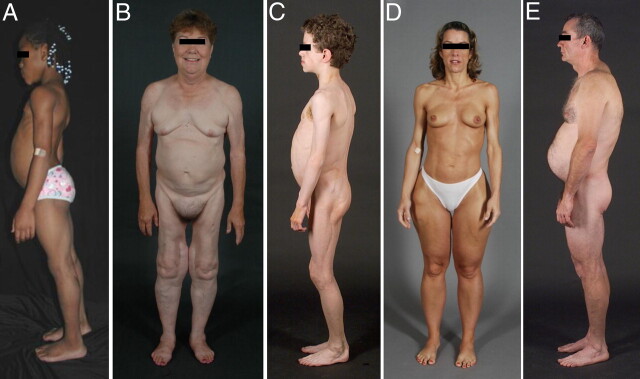
Context: Lipodystrophies are heterogeneous, genetic or acquired disorders characterized by selective loss of body fat and predisposition to insulin resistance. The extent of fat loss determines the severity of associated metabolic complications such as diabetes mellitus, hypertriglyceridemia, and hepatic steatosis.

Evidence acquisition and synthesis: Both original and review articles were found via PubMed search reporting on clinical features and management of various types of lipodystrophies and were integrated with the author's knowledge of the field.

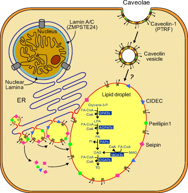
Conclusion: The autosomal recessive congenital generalized lipodystrophy and autosomal dominant familial partial lipodystrophy (FPL) are the two most common types of genetic lipodystrophies. Mutations in AGPAT2, BSCL2, CAV1, and PTRF have been reported in congenital generalized lipodystrophy and in LMNA, PPARG, AKT2, and PLIN1 in FPL. CIDEC is the disease gene for autosomal recessive, FPL and LMNA and ZMPSTE24 for autosomal recessive, mandibuloacral dysplasia-associated lipodystrophy. Recently, an autosomal recessive autoinflammatory lipodystrophy syndrome was reported to be due to PSMB8 mutation. Molecular genetic bases of many rare forms of genetic lipodystrophies remain to be elucidated. The most prevalent subtype of acquired lipodystrophy currently occurs with prolonged duration of protease inhibitor-containing, highly-active antiretroviral therapy in HIV-infected patients. The acquired generalized and partial lipodystrophies are mainly autoimmune in origin and display complement abnormalities. Localized lipodystrophies occur due to drug or vaccine injections, pressure, panniculitis, and other unknown reasons. The current management includes cosmetic surgery and early identification and treatment of metabolic and other complications with diet, exercise, hypoglycemic drugs, and lipid-lowering agents.


Figures

Fig. 1
Fig. 1
Clinical features of patients with various types of lipodystrophies. A, Lateral view of an 8-yr-old African-American female with CGL (also known as Berardinelli-Seip congenital lipodystrophy), type 1 due to homozygous c.377insT (p.Pro128AlafsX19) mutation in AGPAT2. The patient had generalized loss of sc fat with mild acanthosis nigricans in the axillae and neck. She had umbilical prominence and acromegaloid features (enlarged mandible, hands, and feet). B, Anterior view of a 65-yr-old Caucasian female with FPL of the Dunnigan variety due to heterozygous p.Arg482Gln mutation in LMNA. She had marked loss of sc fat from the limbs and anterior truncal region. The breasts were atrophic. She had increased sc fat deposits in the face, anterior neck, suprapubic and vulvar region, and medial parts of the knees. C, Lateral view of an 8-yr-old German boy with AGL. He started experiencing generalized loss of sc fat at age 3 with marked acanthosis nigricans in the neck, axillae, and groin. He developed Crohn's disease at age 11, requiring hemicolectomy at age 13. D, Anterior view of a 39-yr-old Caucasian female with APL (Barraquer-Simons syndrome). She had marked loss of sc fat from the face, neck, upper extremities, chest, and abdomen but had increased sc fat deposition in the lower extremities. E, Lateral view of a 39-yr-old Caucasian male infected with HIV with PI-containing HAART-induced lipodystrophy. He had marked loss of sc fat from the face and limbs but had increased sc fat deposition in the neck region, anteriorly and posteriorly showing buffalo hump. Abdomen was protuberant due to excess intraabdominal fat. He had been on PI-containing antiretroviral therapy for more than 7 yr. [Panel A was reproduced from V. Simha and A. Garg: Lipodystrophy: lessons in lipid and energy metabolism. Curr Opin Lipidol 17:162–169, 2006 (94), with permission. © Lippincott Williams & Williams.]
Fig. 2
Fig. 2
Lipid droplet formation in adipocytes. Lipid droplets (LD) are organelles that store triglycerides (TG) intracellularly. They form as budding vesicles at the endoplasmic reticulum (ER) that fuse in adipocytes to form one large LD. Many proteins, such as CIDEC (shown in blue triangles), seipin (pink squares), and perilipin 1 (green circles) are present on the LD membrane. CIDEC and seipin may be involved in fusion of LDs to form a larger LD, whereas perilipin 1 is essential for lipid storage and hormone-mediated lipolysis. Caveolae are formed from lipid rafts on the cell surface, which include cholesterol (yellow symbols), glycosphingolipids (green symbols), and caveolin-1 (black hairpin-like symbols). Endocytosis of caveolae forms caveolin vesicles that may directly merge with lipid droplets and thus translocating fatty acids to LDs. PTRF controls expression of caveolin 1 and 3 (data not shown). The classical and alternative pathways involved in the biosynthesis of TG are shown inside the lipid droplet. In the adipose tissue, TG synthesis requires glycerol-3-phosphate as the initial substrate (classical pathway), whereas in the small intestine, synthesis of TG can occur via an alternative pathway using monoacylglycerol (MAG) as the initial substrate. Acylation of glycerol-3-phosphate using fatty acyl coenzyme A (FA-CoA) at the sn-1 position is catalyzed by glycerol-3-phosphate acyltransferases (GPATs), resulting in the formation of 1-acylglycerol-3-phosphate or lysophosphatidic acid (LPA). LPA is then acylated at the sn-2 position by AGPATs to yield phosphatidic acid (PA). Removal of phosphate group from PA by PA phosphatases (PAP) produces diacylglycerol (DAG). Further acylation of DAG at the sn-3 position by diacylglycerol acyltransferases (DGATs) finally produces TG. In the alternative pathway, MAG is acylated to DAG by monoacylglycerol acyltransferases (MGATs) which is then further converted to TG. Lamin A/C are integral components of nuclear lamina (shown in blue color) and interact with nuclear membrane proteins as well as chromatin. Zinc metalloproteinase (ZMPSTE24) is critical for posttranslational processing of prelamin A to its mature form, lamin A. [Modified from A. Garg and A. K. Agarwal: Caveolin-1, a new locus for human lipodystrophy. J Clin Endocrinol Metab 93:1183–1185, 2008 (26), with permission. © The Endocrine Society. And from A. Garg and A. K. Agarwal: Lipodystrophies: disorders of adipose tissue biology. Biochem Biophys Acta 1791:507–513, 2009 (95), with permission. © Elsevier.]
Fig. 3
Fig. 3
Pathways involved in the development, differentiation, and death of adipocytes. The pluripotent mesenchymal stem cells can form preadipocytes, myocytes, or osteoblasts depending upon the various cues. In response to various signals from hormones such as insulin and steroids and induction of adipogenic transcription factors, a series of changes are initiated in preadipocytes that lead to their differentiation to adipocytes. The transcription factors, CCAAT (cytidine-cytidine-adenosine-adenosine-thymidine)-enhancer-binding proteins (C/EBP) β/δ are the first to be up-regulated and then stimulate other transcription factors such as PPARγ, C/EBPα, and sterol regulatory element-binding protein (SREBP) 1c. Some other genes such as preadipocyte factor 1 (Pref1), a known adipogenesis inhibitor, are down-regulated. Mature adipocytes are activated, resulting in the overexpression of lipogenic genes like fatty acid synthase (FAS), acetyl coenzyme A carboxylase (ACC), GPAT, AGPAT, and diacylglycerol acyltransferases (DGAT) for biosynthesis of triglycerides and phospholipids. The size of the lipid droplets is reduced upon fasting and increases with increased substrate availability. Available data suggest that the BSCL2-encoded protein, seipin, and AKT2 may be involved in adipocyte differentiation, whereas the AGPAT2 affects triglyceride synthesis. Clinical evidence from lipodystrophy patients harboring LMNA or ZMPSTE24 mutations suggests that nuclear dysfunction may accelerate apoptosis/death of mature adipocytes. Interstitial tissue may also play an important role in adipocyte survival. Mutations in PSMB8, which encodes β5i, a catalytic subunit of the immunoproteasomes, may induce autoinflammatory syndrome, resulting in infiltration of lymphocytes in adipose tissue (panniculitis) and death of nearby adipocytes. [Modified from A. K. Agarwal and A. Garg: Genetic disorders of adipose tissue development, differentiation and death. Annu Rev Genomics Hum Genet 7:175–199, 2006 (96), with permission. © Annual Reviews.]

Similar articles

  • Exploring the pathophysiology behind the more common genetic and acquired lipodystrophies.
    Nolis T. Nolis T. J Hum Genet. 2014 Jan;59(1):16-23. doi: 10.1038/jhg.2013.107. Epub 2013 Oct 24. J Hum Genet. 2014. PMID: 24152769 Review.
  • Lipodystrophies.
    Garg A. Garg A. Am J Med. 2000 Feb;108(2):143-52. doi: 10.1016/s0002-9343(99)00414-3. Am J Med. 2000. PMID: 11126308 Review.
  • [Primary lipodystrophies].
    Capeau J, Magré J, Lascols O, Caron M, Béréziat V, Vigouroux C. Capeau J, et al. Ann Endocrinol (Paris). 2007 Feb;68(1):10-20. doi: 10.1016/j.ando.2006.12.003. Epub 2007 Feb 21. Ann Endocrinol (Paris). 2007. PMID: 17320032 French.
  • Genetic basis of lipodystrophies and management of metabolic complications.
    Agarwal AK, Garg A. Agarwal AK, et al. Annu Rev Med. 2006;57:297-311. doi: 10.1146/annurev.med.57.022605.114424. Annu Rev Med. 2006. PMID: 16409151 Review.
  • Monogenic Forms of Diabetes.
    Sperling MA, Garg A. Sperling MA, et al. In: Cowie CC, Casagrande SS, Menke A, Cissell MA, Eberhardt MS, Meigs JB, Gregg EW, Knowler WC, Barrett-Connor E, Becker DJ, Brancati FL, Boyko EJ, Herman WH, Howard BV, Narayan KMV, Rewers M, Fradkin JE, editors. Diabetes in America. 3rd edition. Bethesda (MD): National Institute of Diabetes and Digestive and Kidney Diseases (US); 2018 Aug. CHAPTER 7. In: Cowie CC, Casagrande SS, Menke A, Cissell MA, Eberhardt MS, Meigs JB, Gregg EW, Knowler WC, Barrett-Connor E, Becker DJ, Brancati FL, Boyko EJ, Herman WH, Howard BV, Narayan KMV, Rewers M, Fradkin JE, editors. Diabetes in America. 3rd edition. Bethesda (MD): National Institute of Diabetes and Digestive and Kidney Diseases (US); 2018 Aug. CHAPTER 7. PMID: 33651552 Free Books & Documents. Review.

Cited by

References

    1. Garg A. 2000. Lipodystrophies. Am J Med 108:143–152 - PubMed
    1. Garg A. 2004. Acquired and inherited lipodystrophies. N Engl J Med 350:1220–1234 - PubMed
    1. Mitchell SW. 1885. Singular case of absence of adipose matter in the upper half of the body. Am J Med Sci 90:105–106
    1. Ziegler LH. 1928. Lipodystrophies: report of seven cases. Brain 51:145–167
    1. Berardinelli W. 1954. An undiagnosed endocrinometabolic syndrome: report of 2 cases. J Clin Endocrinol Metab 14:193–204 - PubMed