. 2011 Nov;85(21):11183-95.
doi: 10.1128/JVI.05468-11. Epub 2011 Aug 24.

Inhibition of dengue virus by targeting viral NS4B protein

Affiliations

Inhibition of dengue virus by targeting viral NS4B protein

Xuping Xie et al. J Virol. 2011 Nov.

Abstract

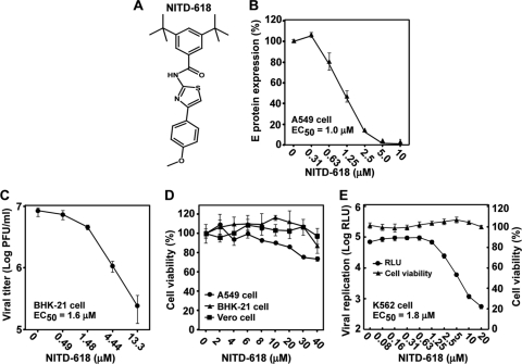
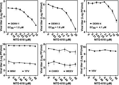
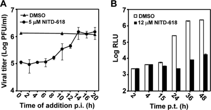
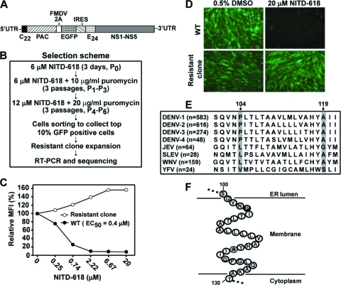
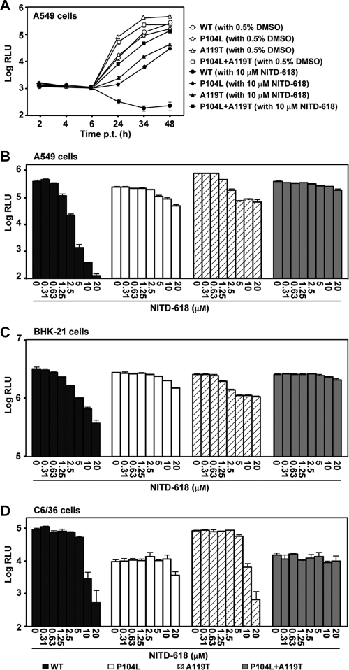
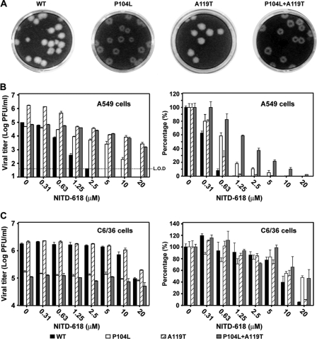
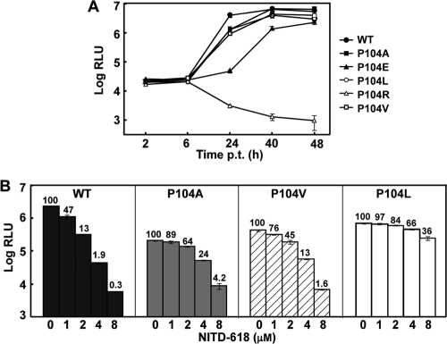
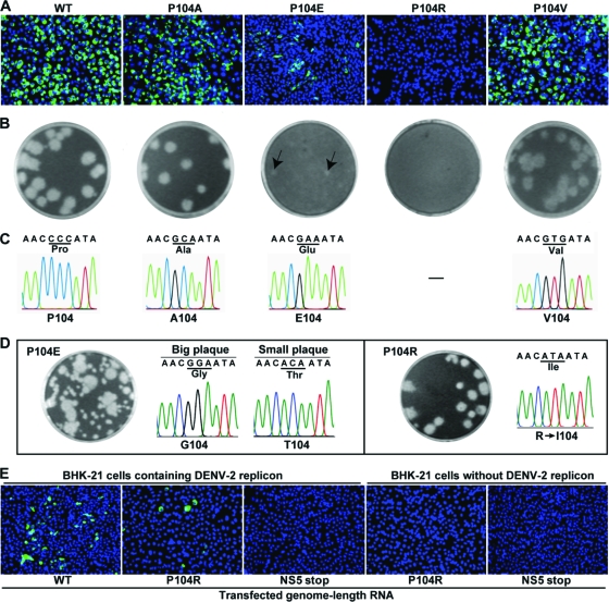
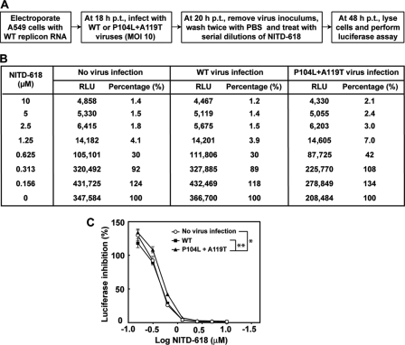
We report a novel inhibitor that selectively suppresses dengue virus (DENV) by targeting viral NS4B protein. The inhibitor was identified by screening a 1.8-million-compound library using a luciferase replicon of DENV serotype 2 (DENV-2). The compound specifically inhibits all four serotypes of DENV (50% effective concentration [EC(50)], 1 to 4 μM; and 50% cytotoxic concentration [CC(50)], >40 μM), but it does not inhibit closely related flaviviruses (West Nile virus and yellow fever virus) or nonflaviviruses (Western equine encephalomyelitis virus, Chikungunya virus, and vesicular stomatitis virus). A mode-of-action study suggested that the compound inhibits viral RNA synthesis. Replicons resistant to the inhibitor were selected in cell culture. Sequencing of the resistant replicons revealed two mutations (P104L and A119T) in the viral NS4B protein. Genetic analysis, using DENV-2 replicon and recombinant viruses, demonstrated that each of the two NS4B mutations alone confers partial resistance and double mutations confer additive resistance to the inhibitor in mammalian cells. In addition, we found that a replication defect caused by a lethal NS4B mutation could be partially rescued through trans complementation. The ability to complement NS4B in trans affected drug sensitivity when a single cell was coinfected with drug-sensitive and drug-resistant viruses. Mechanistically, NS4B was previously shown to interact with the viral NS3 helicase domain; one of the two NS4B mutations recovered in our resistance analysis-P104L-abolished the NS3-NS4B interaction (I. Umareddy, A. Chao, A. Sampath, F. Gu, and S. G. Vasudevan, J. Gen. Virol. 87:2605-2614, 2006). Collectively, the results suggest that the identified inhibitor targets the DENV NS4B protein, leading to a defect in viral RNA synthesis.


Figures

Fig. 1.
Fig. 1.
Antiviral activity of NITD-618 against DENV-2. (A) Structure of NITD-618. (B) Effect of NITD-618 on the expression of DENV-2 E protein. A549 cells were infected with DENV-2 (New Guinea C strain; MOI, 0.3) in the presence of 2-fold serial dilutions of NITD-618. After incubation at 37°C for 48 h, the expression of viral E protein was quantified by CFI assay. The E protein expression level is represented as a percentage of the E expression level derived from the infected cells with DMSO treatment. Each data point shows the average and standard deviation (n = 4). (C) Effect of NITD-618 on the growth of DENV-2. BHK-21 cells were infected with DENV-2 (New Guinea C strain; MOI, 0.3). After incubation at 37°C for 48 h, cell culture fluids were harvested for plaque assay in BHK-21 cells. The curve is reported as logarithm (log10) values of average viral titers of triplicates versus the NITD-618 concentration. The error bars represent standard deviations (n = 3). (D) Cytotoxicity of NITD-618. Cytotoxicity was examined by incubation of A549, BHK-21, and Vero cells with the indicated concentrations of NITD-618. After 48 h of incubation, cell viability was determined with a CCK-8 kit and plotted as a percentage of 0.9% DMSO-treated cells. The average results and standard deviations (n = 3) are presented. (E) Antiviral activity of NITD-618 in K562 cells. K562 cells were infected with a Renilla luciferase DENV-1 (MOI, 1.0) in the presence of NITD-618. At 48 h p.i., the cells were measured for Renilla luciferase activity (relative light units [RLU]) to indicate viral replication and the for intracellular level of ATP to indicate cytotoxicity. The average results and standard deviations from quadruplicate data points are presented.
Fig. 2.
Fig. 2.
Antiviral spectrum of NITD-618. BHK-21 cells were infected with DENV-1, DENV-3, and DENV-4 (MOI, 0.3). A549 cells were infected with CHIKV (MOI, 0.1). Vero cells were infected with WNV, YFV, WEEV, and VSV (MOI, 0.1). EC50s were calculated by nonlinear regression analysis using Prism 5 software. The log10 values of average viral titers and standard deviations (n = 3) are presented.
Fig. 3.
Fig. 3.
Mode-of-action analysis. (A) Time-of-addition analysis. A549 cells were infected with DENV-2 (New Guinea C strain) at an MOI of 2 at 4°C for 1 h. After three washes with PBS, NITD-618 (5 μM) was added to the infected cells at the indicated time points postinfection. As controls, the infected cells were treated with 0.9% DMSO. At 24 h p.i., the culture fluids were harvested, and viral titers were measured by plaque assay. Average results and standard errors (n = 4) are presented. (B) Transient-transfection assay using a luciferase replicon of DENV-2. A549 cells were transfected with equal amounts of WT and mutant replicon RNAs and immediately treated with 12 μM NITD-618 or 0.9% DMSO (negative control). At the indicated time point p.t., cells were assayed for luciferase signals (RLU). The log10 values of average Renilla luciferase signals (RLU) and standard deviations are presented (n = 4).
Fig. 4.
Fig. 4.
Selection and characterization of NITD-618-resistant DENV-2 replicon cells. (A) Schematic diagram of EGFP-expressing DENV-2 replicon (New Guinea C strain). The diagram is not drawn to scale. C22, N-terminal 22 amino acids of the capsid protein; E24, C-terminal 24 amino acids of the E protein; PAC, puromycin acetyltransferase; FMDV 2A, foot-and-mouth disease virus 2A protein; IRES, internal ribosomal entry site from encephalomyocarditis virus. (B) Scheme for selection of resistant replicon cells. See Materials and Methods for details. (C) Resistance analysis of a selected replicon cell line (clone 1). WT and mutant replicon cells were incubated with the indicated concentrations of NITD-618. At 72 h after treatment, the cells were trypsinized, resuspended in PBS containing 2% FBS, and subjected to FACS analysis. The curves were plotted from the mean fluorescence intensity (MFI) versus the concentration of NITD-618. (D) Microscopic analysis of resistance replicon cells. WT and resistant (clone 1) EGFP replicon cells were incubated with 20 μM NITD-618 or 0.5% DMSO (control) for 72 h in the absence of puromycin. Representative microscope images are presented, with EGFP signal in green. (E) Amino acid sequence alignment of NS4B regions. The numbers in parentheses are the numbers of NS4B sequences that were used for the alignment. Sequences were downloaded from the National Center for Biotechnology Information (NCBI) protein database, and alignment was performed using CLC main workbench software (CLC bio). The diagram shows alignment results from amino acid positions 100 to 121 of NS4B. The positions of amino acids in NS4B are numbered according DENV-2 (GenBank accession number AY037116). The identified mutation sites (positions 104 and 119 in DENV-2 NS4B) are shaded. (F) Cartoon diagram of DENV-2 NS4B transmembrane domain 3 (TMD3). The amino acid sequence of TMD3 (residues 100 to 130) of DENV-2 NS4B is shown according to the topology proposed by Miller and colleagues (21). The Pro104 and Ala119 residues identified in this study are highlighted in black and gray, respectively.
Fig. 5.
Fig. 5.
Resistance analysis using a luciferase replicon of DENV-2. (A) A549 cells were transfected with equal amounts (10 μg) of WT, P104L, A119T, or P104L plus A119T mutant replicon RNAs. The transfected cells were immediately treated with NITD-618 (12 μM) or DMSO (0.5%, as a control). Renilla luciferase activities (RLU) were measured at the indicated time points posttransfection. Each data point is a log10 value of the average of luciferase signals from three independent experiments; the error bars indicate standard deviations. (B to D) A549 (B), BHK-21 (C), and C6/36 (D) cells were transfected with WT or mutant replicon RNAs (10 μg). The transfected cells were treated with the indicated concentrations of NITD-618. Luciferase activities were measured at 48 h (A549 and BHK-21 cells) or 96 h (C6/36 cells) posttransfection. Average results and standard deviations from three independent experiments are presented.
Fig. 6.
Fig. 6.
Resistance analysis using recombinant viruses. (A) Plaque morphologies of WT and P104L, A119T, and P104L plus A119T mutant viruses. The plaques were developed in BHK-21 cells (5 days p.i.) without treatment with NITD-618. (B) Viral-titer reduction assay in A549 cells. The dotted line indicates the limit of detection (L.O.D.) of 40 PFU/ml. (C) Viral-titer reduction assay in C6/36 cells. In panels B and C, the log10 values of viral titers versus compound concentrations are plotted on the left, and the percentages of viral titers from the compound-treated samples versus the viral titers from the DMSO-treated samples are shown on the right; for each replicon, the viral titers from the DMSO-treated samples were set as 100%. Average results and standard deviations (n = 3) are presented.
Fig. 7.
Fig. 7.
Comparison of DENV-2 replicons containing different substitutions of NS4B P104. (A) Transient replicon transfection assay. BHK-21 cells were electroporated with equal amounts (10 μg) of WT or NS4B mutant (P104A, P104E, P104L, P104R, or P104V) luciferase replicon RNA. After electroporation, the cells were seeded in 12-well plates (2 × 10⁵ cells/well). At the indicated time point p.t., cells were collected, lysed, and assayed for luciferase activity. Each data point represents the log10 value of the average, and the error bars show the standard deviations (n = 3). (B) Resistance analysis of various replicon mutants. A549 cells were transfected with equal amounts (10 μg) of WT or NS4B mutant (P104A, P104V, or P104L) replicon RNA. The transfected cells were seeded in a 96-well plate (2 × 10⁴ cells/well) and immediately treated with serial dilutions of NITD-618 or 0.5% DMSO (as a control). At 48 h p.t., the cells were washed and lysed, and the luciferase signals were quantified. The data are shown as averages and standard deviations from triplicates. The number above each bar shows the percentage of luciferase units in the treated compared to the untreated samples.
Fig. 8.
Fig. 8.
Analysis of various NS4B P104 mutant viruses. (A) BHK-21 cells were transfected with equal amounts (10 μg) of WT or NS4B mutant genome length RNA of DENV-2. The transfected cells were monitored for viral E protein expression by IFA at 96 h p.t. Anti-E monoclonal antibody 4G2 and Alexa Fluor 488 goat anti-mouse IgG were used as primary and secondary antibodies, respectively. (B) Plaque morphology. Plaques were derived from specific infectivity assays (see Materials and Methods for details). Small plaques of the P104E mutant are indicated by the arrows. (C) Sequencing chromatogram of the mutated NS4B region. Virus RNA recovered from transfected cell supernatant on day 5 p.t. was sequenced for WT, P104A, P104E, and P104V. (D) Revertant analysis of P104E and P104R mutants. For mutant P104E, supernatant from the transfected cells was passaged on Vero cells for six rounds (4 days per round). The passaged viruses yielded mixed plaque morphologies. Plaque purification was performed to isolate viruses exhibiting big plaques and viruses exhibiting small plaques (not shown), and sequencing chromatograms of the plaque-purified viruses are presented. For mutant P104R, supernatant from the transfected cells was passaged on Vero cells for 4 days, and the plaque morphology and sequencing chromatogram of the recovered virus are shown. (E) Trans complementation of the NS4B P104R lethal mutant. BHK-21 cells with or without DENV-2 replicon were transfected with equal amounts (10 μg) of genome length RNA of the WT, NS4B P104R mutant, or NS5stop mutant. The first amino acid (Gly) of NS5 was changed to a UAG stop codon in the NS5stop mutant. At 96 h p.t., the transfected cells were assayed for viral E protein expression using IFA.
Fig. 9.
Fig. 9.
Analysis of trans complementation of resistance. (A) Experimental scheme for trans complementation of resistance. (B) Luciferase activity from the trans complementation experiment. Average luciferase activities (RLU) from quadruplicates are presented. The luciferase activity percentage is equal to the RLU derived from compound-treated sample divided by the RLU derived from the mock-treated control (0.5% DMSO) times 100. (C) Dose-response curve. Prism 5 software was used to plot the data from panel D. The y axis indicates the percent luciferase activity. The x axis indicates the log10 value of the compound concentration. The error bars represent standard deviations (n = 4). The asterisks indicate that the differences are statistically significant (*, P < 0.05; **, P < 0.0001) using a two-way ANOVA test.

Similar articles

Cited by

References

    1. Ackermann M., Padmanabhan R. 2001. De novo synthesis of RNA by the dengue virus RNA-dependent RNA polymerase exhibits temperature dependence at the initiation but not elongation phase. J. Biol. Chem. 276:39926–39937 - PubMed
    1. Bodenreider C., et al. 2009. A fluorescence quenching assay to discriminate between specific and nonspecific inhibitors of dengue virus protease. Anal. Biochem. 395:195–204 - PubMed
    1. Chambers T. J., Grakoui A., Rice C. M. 1991. Processing of the yellow fever virus nonstructural polyprotein: a catalytically active NS3 proteinase domain and NS2B are required for cleavages at dibasic sites. J. Virol. 65:6042–6050 - PMC - PubMed
    1. Chung K. Y., et al. 2010. Higher catalytic efficiency of N-7-methylation is responsible for processive N-7 and 2′-O methyltransferase activity in dengue virus. Virology 402:52–60 - PubMed
    1. Egloff M. P., Benarroch D., Selisko B., Romette J. L., Canard B. 2002. An RNA cap (nucleoside-2′-O-)-methyltransferase in the flavivirus RNA polymerase NS5: crystal structure and functional characterization. EMBO J. 21:2757–2768 - PMC - PubMed

MeSH terms

LinkOut - more resources