. 2011 Nov;85(21):11196-207.
doi: 10.1128/JVI.05601-11. Epub 2011 Aug 24.

Dynamic antibody specificities and virion concentrations in circulating immune complexes in acute to chronic HIV-1 infection

Affiliations

Dynamic antibody specificities and virion concentrations in circulating immune complexes in acute to chronic HIV-1 infection

Pinghuang Liu et al. J Virol. 2011 Nov.

Abstract

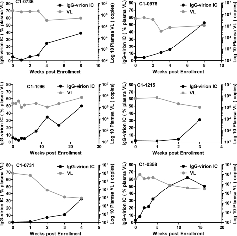
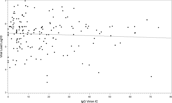
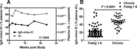
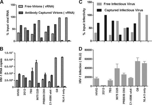
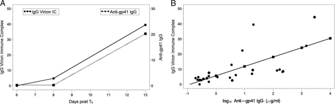
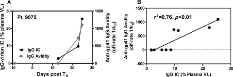
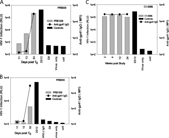
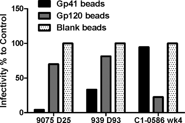
Understanding the interactions between human immunodeficiency virus type 1 (HIV-1) virions and antibodies (Ab) produced during acute HIV-1 infection (AHI) is critical for defining antibody antiviral capabilities. Antibodies that bind virions may prevent transmission by neutralization of virus or mechanically prevent HIV-1 migration through mucosal layers. In this study, we quantified circulating HIV-1 virion-immune complexes (ICs), present in approximately 90% of AHI subjects, and compared the levels and antibody specificity to those in chronic infection. Circulating HIV-1 virions coated with IgG (immune complexes) were in significantly lower levels relative to the viral load in acute infection than in chronic HIV-1 infection. The specificities of the antibodies in the immune complexes differed between acute and chronic infection (anti-gp41 Ab in acute infection and anti-gp120 in chronic infection), potentially suggesting different roles in immunopathogenesis for complexes arising at different stages of infection. We also determined the ability of circulating IgG from AHI to bind infectious versus noninfectious virions. Similar to a nonneutralizing anti-gp41 monoclonal antibody (MAb), purified plasma IgG from acute HIV-1 subjects bound both infectious and noninfectious virions. This was in contrast to the neutralizing antibody 2G12 MAb that bound predominantly infectious virions. Moreover, the initial antibody response captured acute HIV-1 virions without selection for different HIV-1 envelope sequences. In total, this study demonstrates that the composition of immune complexes are dynamic over the course of HIV-1 infection and are comprised initially of antibodies that nonselectively opsonize both infectious and noninfectious virions, likely contributing to the lack of efficacy of the antibody response during acute infection.


Figures

Fig. 1.
Fig. 1.
Increasing proportion of acute HIV-1 plasma virions are coated by IgG. Endogenous plasma IgG-virion immune complexes in plasma from 38 AHI subjects were measured by the use of protein G absorption. The IgG-virion immune complexes were detected in the plasma of 34 out of 38 subjects (IgG opsonized a range of 8.6 to 73.89% of plasma HIV-1 VL during AHI). Endogenous IgG-virion IC and plasma viral load for 6 representative subjects (enrolled at Fiebig stages I to IV) are shown.
Fig. 2.
Fig. 2.
No correlation between HIV-1 plasma viral load and plasma endogenous IgG-virion IC in all samples from AHI. The plasma viral load was plotted against the level of the endogenous plasma IgG-virion IC (r = 0.06, P = 0.41).
Fig. 3.
Fig. 3.
The plasma endogenous IgG-virion IC in chronic subjects. (A) Unlike AHI subjects, chronic subjects maintained a high level of endogenous IgG-virion IC over time. Approximately 50% plasma viral load was captured by IgG in the chronic subject C1-0645 over a 24-week study period. (B) Comparison of the endogenous IgG-virion IC in acute and chronic infection. The value of endogenous plasma IgG-virion IC in 39 samples from chronic infection (10 chronic subjects) compared to 69 samples from acute infection; Fiebig stages I to VI in 34 AHI subjects are shown here. The Fiebig stage was defined as described in reference . The samples from plasma donor cohort were assigned to Fiebig stages I to VI if they were less than 30 days post-T0. The line bar represents the mean value (P values, unpaired two-tailed Student t test).
Fig. 4.
Fig. 4.
Plasma IgG from HIV-1 infection captures infectious or noninfectious HIV-1 viral particles. IgG-virion IC was prepared in vitro at final concentration of 10 μg/ml IgG, and IgG-virion IC mixture was absorbed by protein G. The viral RNA or infectivity in different fractions was measured by HIV-1 Gag real-time RT-PCR (Fig. 4A and B) or TZM-bl infection (Fig. 4C and D). The error bar represents the standard deviation. The percentage of virions (viral RNA) (Fig. 4A) or infectious viruses in each fraction (Fig. 4C) of the input was calculated as described in Materials and Methods and are shown here. vRNA, viral RNA.
Fig. 5.
Fig. 5.
Circulating plasma IgG-virion immune complexes significantly correlate with the concentrations of anti-gp41 Env IgG during AHI. (A) IgG-virion IC and anti-gp41 IgG from one typical plasma donor subject (patient 9012) is presented here. (B) Levels of log10 anti-gp41 IgG (μg/ml) in plasma significantly correlate with the level of plasma IgG-HIV-1 virion IC (n = 8) (t = 4.88, P = 0.0001). Every log10 increase in anti-gp41 IgG correlates to a 6.2% increase in IgG-virion IC. Each color represents one individual subject.
Fig. 6.
Fig. 6.
Endogenous plasma IgG-virion IC correlates with the dissociation rates (avidity maturation) of plasma anti-gp41 IgG. (A) The anti-gp41 IgG avidity (off rate,1/Kd in seconds) and the endogenous plasma IgG-virion IC are shown for a representative subject during acute infection. (B) Levels of IgG-virion IC and the anti-gp41 IgG dissociation rates significantly correlate (n = 4) (r2 = 0.76, P < 0.01). Dissociation rates (s−1) as a measure of avidity were calculated as described in Materials and Methods.
Fig. 7.
Fig. 7.
Purified plasma IgG from AHI (A, B) or chronic (C) subjects captures infectious HIV-1. A total of 50 μl IC mixture containing plasma IgG at 10 μg/ml and 5 × 10⁶ RNA copies viral particles was added to each mouse anti-human IgG-coated well after incubation in vitro for 1.5 h. The infection of captured IgG IC was measured by multiple-round M7-luc assay for NL-LucR.T2A at 5 days postinfection (A and C) or by TZM-bl at 3 days postinfection for NL-LucR.T2A-WITO (B). The infectivity was measured by firefly luciferase assay and expressed as relative luciferase units (RLU). The gray bar represents the positive or negative controls. The gray bar represents the clinical samples. The levels of anti-gp41 IgG of plasma samples (solid line) were plotted together with the in vitro capture. The cutoff values were 3× background. All runs were performed in triplicate.
Fig. 8.
Fig. 8.
Different specificities of antibodies in immune complexes in acute and chronic infection. Purified plasma IgG from AHI subjects 9075 (day 25 post-T0) and PRB939 (day 93 post-T0) or chronic subject C1-0586 (week 4 poststudy) were absorbed by Env gp41 or Env gp120-conjugated, or blank microsphere beads. Then the absorbed supernatant was used to prepare IgG-virion IC mixture in vitro. The infectious IC was captured by plates coated with anti-human IgG antibody and was measured for infection in the M7-luc infection assay as described above. The percentage of infection compared to the control blank bead is shown here. The results represent at least 3 experiments.
Fig. 9.
Fig. 9.
Percentage of virions bound in complexes at various antibody concentrations for 8 different patients. Red lines show the best fits to patient data for a model with univalent binding (red dots), given by equation 1. Blue and green lines assume HIV has binding sites with f values of 14 and 35, respectively. Estimates for the affinity constant K are given in Table S2 in the supplemental material. The aberrant fit of the model to the data from patient 12008 could be explained by an extension of the model that assumes that two different strains of HIV-1 with different affinities for anti-gp41 IgG are present in this patient (results not shown).

References

    1. Alam S. M., et al. 2008. Human immunodeficiency virus type 1 gp41 antibodies that mask membrane proximal region epitopes: antibody binding kinetics, induction, and potential for regulation in acute infection. J. Virol. 82:115–125 - PMC - PubMed
    1. Bajtay Z., Speth C., Erdei A., Dierich M. P. 2004. Cutting edge: productive HIV-1 infection of dendritic cells via complement receptor type 3 (CR3, CD11b/CD18). J. Immunol. 173:4775–4778 - PubMed
    1. Bouhlal H., et al. 2002. Opsonization of HIV-1 by semen complement enhances infection of human epithelial cells. J. Immunol. 169:3301–3306 - PubMed
    1. Bourinbaiar A. S. 1994. The ratio of defective HIV-1 particles to replication-competent infectious virions. Acta Virol. 38:59–61 - PubMed
    1. Burrer R., Haessig-Einius S., Aubertin A. M., Moog C. 2005. Neutralizing as well as non-neutralizing polyclonal immunoglobulin (Ig)G from infected patients capture HIV-1 via antibodies directed against the principal immunodominant domain of gp41. Virology 333:102–113 - PubMed

Publication types

Substances