. 2011 Aug 24;3(97):97ra81.
doi: 10.1126/scitranslmed.3002473.

Potent kinetic stabilizers that prevent transthyretin-mediated cardiomyocyte proteotoxicity

Affiliations

Potent kinetic stabilizers that prevent transthyretin-mediated cardiomyocyte proteotoxicity

Mamoun M Alhamadsheh et al. Sci Transl Med. .

Abstract

A valine-to-isoleucine mutation at position 122 of the serum protein transthyretin (TTR), found in 3 to 4% of African Americans, alters its stability, leading to amyloidogenesis and cardiomyopathy. In addition, 10 to 15% of individuals older than 65 years develop senile systemic amyloidosis and cardiac TTR deposits because of wild-type TTR amyloidogenesis. Although several drugs are in development, no approved therapies for TTR amyloid cardiomyopathy are yet available, so the identification of additional compounds that prevent amyloid-mediated cardiotoxicity is needed. To this aim, we developed a fluorescence polarization-based high-throughput screen and used it to identify several new chemical scaffolds that target TTR. These compounds were potent kinetic stabilizers of TTR and prevented TTR tetramer dissociation, partial unfolding, and aggregation of both wild type and the most common cardiomyopathy-associated TTR mutant, V122I-TTR. High-resolution co-crystal structures and characterization of the binding energetics revealed how these diverse structures bound to tetrameric TTR. These compounds effectively inhibited the proteotoxicity of V122I-TTR toward human cardiomyocytes. Several of these ligands stabilized TTR in human serum more effectively than diflunisal, which is a well-studied inhibitor of TTR aggregation, and may be promising leads for the treatment or prevention of TTR-mediated cardiomyopathy.


Conflict of interest statement

Competing interests: The authors declare that they have no competing interests.

Figures
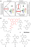
Fig. 1
Fig. 1
Schematic of the FP assay and structures of TTR ligands and FP probe 1. (A) The rate of tumbling of fluorescent probe 1 decreases upon binding to TTR, which results in increasing its fluorescence polarization signal. (B) Chemical structures of the TTR ligands 2–5 used to design probe 1.
Fig. 2
Fig. 2
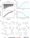
Assessment of FP probe 1 binding to TTR and structures of TTR ligands. (A) Assessment of the binding affinity of probe 1 to TTR by ITC. Calorimetric titration of probe 1 against TTR (Kd,1 = 13 nM and Kd,2 = 100 nM). Raw data (top) and integrated heats (bottom) from the titration of TTR (2 μM) with probe (25 μM). The solid red line represents the best fit binding isotherm to a two-site binding model. (B) FP saturation binding between probe 1 (0.1 μM) and increasing concentration of TTR (0.005 to 10 μM) (C) Competition of probe 1 from TTR by increasing concentrations (0.01 μM – 50 μM) of ligand 2 (Kapp = 0.231 μM, R2 = 0.997). FP Assays were performed in triplicate. Error bars: SD. (D) Structures of the newly identified TTR ligands. Compounds with co-crystal structures in Fig. 5 are labeled in red in all figures.
Fig. 3
Fig. 3
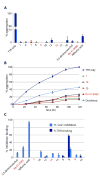
Evaluation of inhibition of TTR aggregation and binding to THR and COX-1 (A) Percentage of TTR (3.6 μM) fibril formation in the presence of ligands (7.2 μM) relative to aggregation in the absence of ligands (denoted 100%) after 72 hours. (B) Comparison of TTR (3.6 μM) aggregation inhibition in the presence of substoichiometric amounts of ligands (3.0 μM) compared to diclofenac. (C) THR binding and COX-1 inhibition data for the most potent TTR ligands. THR binding is expressed as % displacement of 125I-labeled triiodothyronine (T3) by test compounds (10 μM). COX-1 inhibition is shown as % inhibition of COX-1 mediated conversion by test compounds (10 μM). Error bars: SEM
Fig. 4
Fig. 4
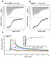
Assessment of the binding of ligands Ro 41-0960 and 7 to TTR using ITC and SPR. Calorimetric titration of (A) Ro 41-0960 (Kd,1 = 15 nM and Kd,2 = 2000 nM) and (B) 7 (Kd,1 = 58 nM and Kd,2 = 500 nM) against TTR. Raw data (top) and integrated heats (bottom) from the titration of TTR (2 μM) with ligand (25 μM). The solid red line through the integrated heats represents the best fit binding isotherm to a two-site binding model. (C) SPR sensogram showing concentration-dependent binding of ligand 7 to TTR (0.001 μM to 2.2 μM). Normalized RUs are plotted over a time course. Equilibrium binding analysis (inset) indicates an apparent Kd of 57.91 ± 13.2 nM (SD).
Fig. 5
Fig. 5
Crystal structures of WT-TTR bound to T4, Ro 41-0960, 14, 7, and 9 (A) Ribbon diagram of WT-TTR with T4 bound to each of the two T4 binding pockets within the tetramer (monomer subunits individually colored). (B) Expanded view of T4 within the binding site with a “Connelly” analytical molecular surface applied to residues within 8 Å of ligand (green = hydrophobic, purple = polar). (C to F) Expanded views of Ro 41-0960, 14, 7, and 9 bound to TTR. Hydrogen bonds are represented as dashed lines between functional groups. The innermost halogen binding pockets (HBPs) 3 and 3′ are composed of the methyl and methylene groups of Ser117/117′, Thr119/119′, and Leu110/110′. HBPs 2 and 2′ are assembled from the side chains of Leu110/110′, Ala109/109′, Lys15/15′, and Leu17/17′. The outermost HBPs 1 and 1′ (labeled in a) are lined by the methyl and methylene groups of Lys15/15′, Ala108/108′, and Thr106/106 ′ (Thr 106 not shown).
Fig. 6
Fig. 6
Inhibition of V1221I TTR cytotoxicity and stabilization of human serum TTR. (A) Inhibition of V1221I TTR cytotoxicity towards AC16 cells. TTR and ligands concentration: 8 μM. Resveratrol is a positive control. Cell viability is shown relative to cells treated with vehicle only. Error bars: SEM (B) Quantitative analysis of TTR serum stability after 72 hours. (C&D) Stabilization of human serum TTR. Serum TTR in the presence and absence 50 μM compounds was subjected to acid denaturation. TTR immunoblot at 0 (C) and 72 (D) hours after acidification of serum

Comment in

Similar articles

Cited by

References

    1. Selkoe DJ. Folding proteins in fatal ways. Nature. 2003;426:900–904. - PubMed
    1. Cohen FE, Kelly JW. Therapeutic approaches to protein-misfolding diseases. Nature. 2003;426:905–909. - PubMed
    1. Johnson SM, et al. Native state kinetic stabilization as a strategy to ameliorate protein misfolding diseases: a focus on the transthyretin amyloidoses. Acc Chem Res. 2005;38:911–921. - PubMed
    1. Monaco HL, Rizzi M, Coda A. Structure of a complex of two plasma proteins: transthyretin and retinol-binding protein. Science. 1995;268:1039–1041. - PubMed
    1. Falk RH, Comenzo RL, Skinner M. The systemic amyloidoses. N Engl J Med. 1997;337:898–909. - PubMed

Publication types