. 2011 Dec;85(6):1279-83.
doi: 10.1095/biolreprod.111.094094. Epub 2011 Aug 24.

Age-dependent susceptibility of chromosome cohesion to premature separase activation in mouse oocytes

Affiliations

Age-dependent susceptibility of chromosome cohesion to premature separase activation in mouse oocytes

Teresa Chiang et al. Biol Reprod. 2011 Dec.

Abstract

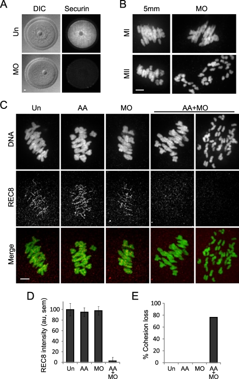
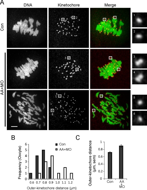
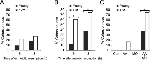
A hypothesis to explain the maternal age-dependent increase in formation of aneuploid eggs is deterioration of chromosome cohesion. Although several lines of evidence are consistent with this hypothesis, whether cohesion is actually reduced in naturally aged oocytes has not been directly tested by any experimental perturbation. To directly target cohesion, we increased the activity of separase, the protease that cleaves the meiotic cohesin REC8, in oocytes. We show that cohesion is more susceptible to premature separase activation in old oocytes than in young oocytes, demonstrating that cohesion is significantly reduced. Furthermore, cohesion is protected by two independent mechanisms that inhibit separase, securin and an inhibitory phosphorylation of separase by CDK1; both mechanisms must be disrupted to prematurely activate separase. With the continual loss of cohesins from chromosomes that occurs throughout the natural reproductive lifespan, tight regulation of separase in oocytes may be particularly important to maintain cohesion and prevent aneuploidy.


Figures

FIG. 1.
FIG. 1.
Cohesion is maintained by two independent mechanisms that inhibit separase. A) Uninjected oocytes (Un; n = 10) or oocytes microinjected with securin MO (n = 17) were fixed and stained for securin protein in germinal vesicle-intact oocytes. B) Oocytes microinjected with securin MO or a 5-bp mismatch MO (5 mm) were matured in vitro to MI (MO, n = 19; 5 mm, n = 11) or MII (MO, n = 10; 5 mm, n = 8), fixed, and stained for DNA. C and D) Un oocytes (n = 23) or oocytes microinjected with AA-separase cRNA (AA; n = 18), securin MO (n = 14), or both (AA+MO; n = 21) were fixed 8 h after meiotic resumption, and REC8 protein was detected by immunocytochemistry. Oocytes were collected from a total of five mice. Representative images (C) are maximal intensity projections of confocal z-series showing DNA (green) and REC8 (red). Note that in AA+MO oocytes, REC8 levels are low even when bivalents are intact. REC8 fluorescence intensity (mean ± SEM) was normalized to the mean intensity of Un oocytes and quantified for each group (D). E) Percentage of oocytes with cohesion loss, as indicated by prematurely separated bivalents and chromatids, was determined for each group. Bar = 5 μm.
FIG. 2.
FIG. 2.
Centromeres are more susceptible than chromosome arms to cohesin loss. A) Control oocytes (Con; n = 12) or oocytes microinjected with both AA-separase cRNA and securin MO (AA+MO; n = 23) were fixed 8 h after meiotic resumption and stained for DNA (green) and kinetochores (CREST; red). The AA+MO group includes oocytes with intact chromosomes (middle; n = 12 of 23) and prematurely separated chromosomes (bottom; n = 11 of 23). Images are maximal intensity projections of confocal z-series, with insets of single optical sections to show kinetochores. Insets 1–4 include the two kinetochores of a sister pair; insets 5 and 6 show single kinetochores in AA+MO oocytes where chromosomes prematurely separated. Oocytes were collected from a total of four mice. Bar = 5 μm. B and C) Outer kinetochore distances were measured from the outer edges of sister kinetochore pairs for control and AA+MO oocytes with intact chromosomes (n = 240 kinetochore pairs from 12 oocytes in each group). The populations are represented by histograms (B) and by the mean ± SEM for each group (C).
FIG. 3.
FIG. 3.
Cohesion is reduced in old oocytes. A) Young (n = 22 from four mice) and 12m oocytes (n = 18 from eight mice) microinjected with a cRNA-encoding fluorescently labeled H2B, AA-separase cRNA, and securin MO to increase separase activity were matured in vitro and imaged live by fluorescence at 5 and 8 h after meiotic resumption. Percentage of oocytes with cohesion loss, as indicated by prematurely separated bivalents and chromatids, was determined for each group. At both time points, bivalents in young and 12m oocytes are similarly intact. B) Young and old oocytes were microinjected as in A and imaged at 5 h (n = 17 young and 13 old) and 8 h (n = 29 young and 20 old) after meiotic resumption. Percentage of oocytes with cohesion loss is significantly higher in old oocytes compared to young (asterisks) at both 5 h (Fisher exact test, P = 0.007) and 8 h (P = 0.019). Oocytes were collected from 11 young and 10 old mice. C) Young and old oocytes were microinjected with a cRNA-encoding fluorescently labeled H2B (Control; n = 94 young and 14 old), AA-separase cRNA (AA; n = 6 young and 12 old), securin MO (MO; n = 13 young and 9 old), or both AA and MO (AA+MO; n = 29 young and 20 old) and imaged at 8 h after meiotic resumption. The increased cohesion loss in old AA oocytes is not statistically significant. Oocytes were collected from 34 young and 34 old mice.

References

    1. Hassold T, Hall H, Hunt P. The origin of human aneuploidy: where we have been, where we are going. Hum Mol Genet 2007; 16(spec no. 2): R203 R208 - PubMed
    1. Tachibana-Konwalski K, Godwin J, van der Weyden L, Champion L, Kudo NR, Adams DJ, Nasmyth K. Rec8-containing cohesin maintains bivalents without turnover during the growing phase of mouse oocytes. Genes Dev 2010; 24: 2505 2516 - PMC - PubMed
    1. Revenkova E, Herrmann K, Adelfalk C, Jessberger R. Oocyte cohesin expression restricted to predictyate stages provides full fertility and prevents aneuploidy. Curr Biol 2010; 20: 1529 1533 - PMC - PubMed
    1. Hodges CA, Revenkova E, Jessberger R, Hassold TJ, Hunt PA. SMC1beta-deficient female mice provide evidence that cohesins are a missing link in age-related nondisjunction. Nat Genet 2005; 37: 1351 1355 - PubMed
    1. Subramanian VV, Bickel SE. Aging predisposes oocytes to meiotic nondisjunction when the cohesin subunit SMC1 is reduced. PLoS Genet 2008; 4: e1000263. - PMC - PubMed

Publication types