. 2011 Oct;22(20):3861-73.
doi: 10.1091/mbc.E11-01-0080. Epub 2011 Aug 24.

Ligand activation leads to regulated intramembrane proteolysis of fibroblast growth factor receptor 3

Affiliations

Ligand activation leads to regulated intramembrane proteolysis of fibroblast growth factor receptor 3

Catherine R Degnin et al. Mol Biol Cell. 2011 Oct.

Abstract

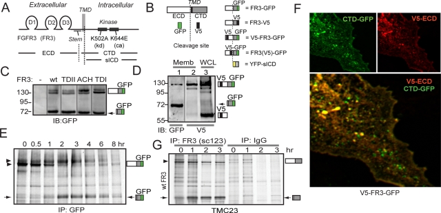
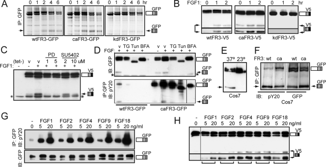
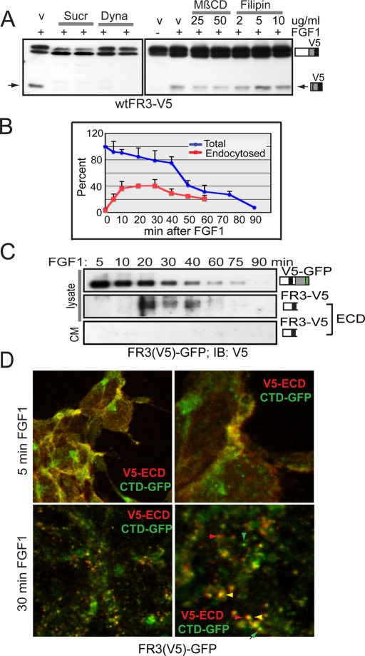
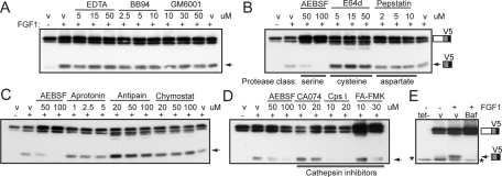
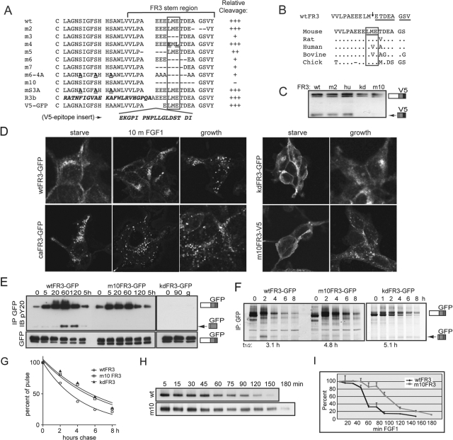
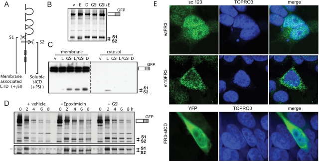
Fibroblast growth factor receptor 3 (FGFR3) is a major negative regulator of bone growth that inhibits the proliferation and differentiation of growth plate chondrocytes. Activating mutations of its c isoform cause dwarfism in humans; somatic mutations can drive oncogenic transformation in multiple myeloma and bladder cancer. How these distinct activities arise is not clear. FGFR3 was previously shown to undergo proteolytic cleavage in the bovine rib growth plate, but this was not explored further. Here, we show that FGF1 induces regulated intramembrane proteolysis (RIP) of FGFR3. The ectodomain is proteolytically cleaved (S1) in response to ligand-induced receptor activation, but unlike most RIP target proteins, it requires endocytosis and does not involve a metalloproteinase. S1 cleavage generates a C-terminal domain fragment that initially remains anchored in the membrane, is phosphorylated, and is spatially distinct from the intact receptor. Ectodomain cleavage is followed by intramembrane cleavage (S2) to generate a soluble intracellular domain that is released into the cytosol and can translocate to the nucleus. We identify the S1 cleavage site and show that γ-secretase mediates the S2 cleavage event. In this way we demonstrate a mechanism for the nuclear localization of FGFR3 in response to ligand activation, which may occur in both development and disease.


Figures

FIGURE 1:
FIGURE 1:
FGFR3 is proteolytically cleaved when expressed in the presence of serum. (A) Cartoon of relevant FGFR3 structures. D1–3: D-loops. CTD, C-terminal domain; ECD, extracellular domain; sICD, soluble intracellular domain; TMD, transmembrane domain. FR3, FGFR3. (B) Cartoon of FGFR3 constructs and epitope tags. (C–G) Arrow, cleaved FR3 CTD. (C) Western blot of control and Cos7 cell lysates stably expressing GFP-tagged wt and mutant FR3, probed for GFP epitope. Upper bands, intact receptor. (D) Membrane extracts (lanes 1, 2) and whole-cell lysates (WCL) (lane 3) prepared from Cos7 cells transiently expressing V5-FR3-GFP, probed for GFP (lane 1) or V5 (lanes 2, 3). (E) Pulse-chase analysis of Cos7 cells stably expressing wtFR3-GFP, immunoprecipitated for the GFP epitope. Arrowheads, mature FR3. (F) Confocal analysis of V5-FR3-GFP transiently expressed in Cos7 cells. Green, C-terminal GFP; red, N-terminal V5; yellow, colocalized epitopes. (G) Endogenous FGFR3 is cleaved. Pulse-chase analysis of TMC-23 cells immunoprecipitated for FGFR3 (sc123) or control immunoglobulin G.
FIGURE 2:
FIGURE 2:
FGF1-induced cleavage requires FGFR3 activation. (A–H) Arrow, cleaved FR3; asterisk, nonspecific band. (A) Pulse-chase analysis of T-Rex 293 cell lines stably expressing wt, ca, or kdFR3-GFP in growth media, immunoprecipitated for GFP epitope. (B–D) Western blots of wtFR3-expressing T-Rex 293 cells, cultured in DMEM/FBS(–) media in the presence or absence of FGF1 and probed for C-terminal V5 epitope, unless stated otherwise. (B) Serum-starved cells activated with FGF1 for the times indicated. Bottom, longer exposure to show that kd receptor is not cleaved. (C) Cells treated 8 h with or without tet/FGF1, ± PD173074 (PD), or SU5402 as indicated. v, vehicle (dimethyl sulfoxide [DMSO]). (D) Cells cultured 8 h in the presence or absence of inhibitor as indicated. BFA, 6 μg/ml brefeldin A; TG, 1 mg/ml thapsigarin; Tun, 1 μg/ml tunicamycin; v, DMSO. Top, 1/10 WCL, probed for GFP. Bottom, IP GFP, IB phosphotyrosine (pY20). wtFR3, left; caFR3, right. (E) Western blot of Cos7 cells stably expressing wtFR3-GFP cultured 5 h at 37°C (left) or 23°C (right), probed for GFP. (F) Western blot of Cos7 cells stably expressing wt or caFR3-GFP cultured under growth conditions, immunoprecipitated for GFP and probed for phosphotyrosine (pY20, left). WCL 1/10 probed for GFP (right). (G) Serum-starved wtFR3-GFP expressing T-Rex 293 cells, activated 5 min with the growth factors indicated. Equal micrograms of lysate were immunoprecipitated for GFP and probed for phosphotyrosine (pY20). WCL 1/10 was probed for GFP (bottom, IB GFP). (H) T-Rex 293 cells expressing wtFR3 were tet induced for 8 h in DMEM/FBS(−) media containing the FGF species indicated. Western blot of lysates probed with V5 antibodies. Bottom, longer exposure to show relative amounts of cleaved FR3.
FIGURE 3:
FIGURE 3:
Endocytosis is required for FGF1-induced cleavage of FGFR3. (A) Western blot of lysates from wtFR3-V5–expressing T-Rex 293 cells induced 3 h ± FGF1 before treating an additional 5 h with drug or DMSO (v). Dyna, 67, 80 μM dynasore; MβCD, methyl-β-cyclodextrin; Sucr, 0.4 M sucrose. (B) Graphical analysis of three independent experiments plotting loss of intact cell surface biotinylated receptor (blue line) and endocytosis of intact, cell surface biotinylated, glutathione-resistant receptor (red line). SDs are given as error bars. (C) FR3(V5)-GFP expressing cells were serum starved, cell surface biotinylated, and then treated with FGF1 for the times indicated. Equal micrograms of lysate or all the culture media (CM) was affinity purified using NeutrAvidin Gel. Western blot using equal volumes of purified lysate or one half of purified CM was probed for N-terminal V5 epitope (ECD). ECD was detected by overexposing blots. (D) Serum-starved FR3(V5)-GFP–expressing cells were surface labeled with V5 antibodies, then stimulated with FGF1 to promote receptor internalization. Cells were fixed, permeabilized, probed with secondary antibodies (Alexa 543) to the endocytosed V5 epitope, and imaged by confocal microscopy. Right, higher-magnification image of left. Green, C-terminal GFP; red, endocytosed ECD-V5. Green arrowheads, GFP-containing vesicles; red arrowheads, ECD-containing vesicles; yellow arrowhead, colocalized ECD/CTD.
FIGURE 4:
FIGURE 4:
FGFR3 cleavage involves endosomal cathepsins. (A–E) wtFR3-V5–expressing T-Rex 293 cells treated with tet/FGF1, ± inhibitor, or DMSO (v), as indicated. Western blot of lysates probed for the V5 epitope. Arrow, cleaved FR3; asterisk, nonspecific band. Baf, 250 μM bafilomycin A1; Chymostat, chymostatin; Cps I, cathepsin inhibitor I.
FIGURE 5:
FIGURE 5:
Disrupting cleavage alters the half-life, but not the trafficking, of FGFR3. (A–I) T-Rex 293 stable cell lines. Arrow, cleaved FR3. (A) Stem region sequence of mutants (m) generated and relative degree of cleavage, averaged from at least three independent experiments. Cell lines were tet-induced 8 h in the presence of FGF1; cleavage was assessed by Western blot using V5 or GFP epitope. +++, wt cleavage; ++, reduced cleavage; +, minimal cleavage; −, no cleavage detected. Putative cleavage region is boxed. Mutated residues are shown in bold; FR3b-specific sequence and V5 epitope insertion are shown in bold and italicized. (B) Top, cleavage site determined by N-terminal sequencing of gel-purified 72-kDa fragment. Arrow, cleavage site. Sequenced residues are underlined. Bottom, homology of cleavage region between species. Residues surrounding cleavage site are boxed; distinct amino acids are listed. (C) Representative Western blot of various stable FR3-V5 cell lines probed for V5 epitope. hu, human stem region. (D) Confocal images of wt and mutant FR3 following serum starvation, 10 min FGF1 addition, or cultured in growth media. GFP imaged directly; V5 probed with V5 antibody and Alexa 488 secondary antibody. (E) Cells expressing wt and mutant FR3-GFP were serum starved and then treated with FGF1 for the times indicated. Equal micrograms of lysates were immunoprecipitated for GFP and probed for phosphotyrosine (pY20). (F) Cells expressing wt, m10, and kdFR3-GFP were subject to pulse-chase analysis in the presence of FGF1 and immunoprecipitated for GFP. Arrow, cleaved FR3. T1/2, half-life of intact receptor calculated using GraphPad (La Jolla, CA). (G) Graphical depiction of the half-life for intact wt, m10, and kdFR3. (H) Cells expressing wt or m10FR3-V5 were serum starved, cell surface biotinylated, and stimulated with FGF1 for the times indicated. Equal micrograms of lysate were immunoprecipitated using NeutrAvidin Gel; the stability of the intact receptor was assessed by Western blot using the V5 epitope. (I) Graphical analysis of three independent experiments assaying loss of intact cell surface biotinylated receptor. SDs are shown as error bars.
FIGURE 6:
FIGURE 6:
FGFR3 is cleaved by γ-secretase. (A) Cartoon of cleavage sites (S1, putative cathepsin; S2, γ-secretase) and their subcellular localization. γSI, γ-secretase inhibitor; PSI, proteasome inhibitor. (A) Cells expressing wtFR3-GFP were pulse labeled in the presence of FGF1 and then chased 2.5 h in the presence of inhibitors. D, 25 μM DAPT; E, 1 μM epoxomicin; GSI, 5 μM compound E; v, DMSO. (C) Western blot of cells expressing wtFR3-GFP, cultured 5 h in the presence of inhibitors, subject to subcellular fractionation, and probed for C-terminal GFP tag. Left, membrane-associated proteins; right, cytosolic proteins. L, 10 μM lactacystin. (D) Cells expressing wtFR3-GFP were pulse labeled and then chased in the presence of FGF1 plus inhibitors. Bottom, longer exposure to show the size shift of the cleaved fragment. Dashed line, S1-cleaved FR3. (E) Confocal images of cells transiently overexpressing untagged wtFR3 (top), m10FR3 (middle), or YFP-tagged FR3-sICD (bottom, YFP-sICD). sc 123, C-terminal anti-FGFR3 antibody (green); YFP pseudo-colored green; TOPRO3, nuclear stain (blue).

Similar articles

Cited by

References

    1. Alwan HA, van Zoelen EJ, van Leeuwen JE. Ligand-induced lysosomal epidermal growth factor receptor (EGFR) degradation is preceded by proteasome-dependent EGFR de-ubiquitination. J Biol Chem. 2003;278:35781–35790. - PubMed
    1. Ancot F, Foveau B, Lefebvre J, Leroy C, Tulasne D. Proteolytic cleavages give receptor tyrosine kinases the gift of ubiquity. Oncogene. 2009;28:2185–2195. - PubMed
    1. Bae JH, Boggon TJ, Tome F, Mandiyan V, Lax I, Schlessinger J. Asymmetric receptor contact is required for tyrosine autophosphorylation of fibroblast growth factor receptor in living cells. Proc Natl Acad Sci USA. 2010;107:2866–2871. - PMC - PubMed
    1. Bank U, Reinhold D, Schneemilch C, Kunz D, Synowitz HJ, Ansorge S. Selective proteolytic cleavage of IL-2 receptor and IL-6 receptor ligand binding chains by neutrophil-derived serine proteases at foci of inflammation. J Interferon Cytokine Res. 1999;19:1277–1287. - PubMed
    1. Belleudi F, Leone L, Nobili V, Raffa S, Francescangeli F, Maggio M, Morrone S, Marchese C, Torrisi MR. Keratinocyte growth factor receptor ligands target the receptor to different intracellular pathways. Traffic. 2007;8:1854–1872. - PubMed

Publication types

MeSH terms

Substances