. 2011 Oct;22(20):3749-57.
doi: 10.1091/mbc.E11-04-0293. Epub 2011 Aug 24.

Mia40-dependent oxidation of cysteines in domain I of Ccs1 controls its distribution between mitochondria and the cytosol

Affiliations

Mia40-dependent oxidation of cysteines in domain I of Ccs1 controls its distribution between mitochondria and the cytosol

Christine Klöppel et al. Mol Biol Cell. 2011 Oct.

Abstract

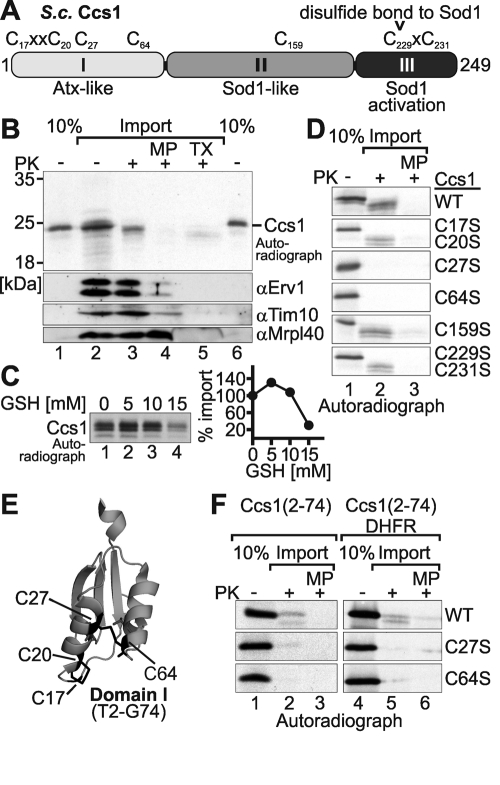
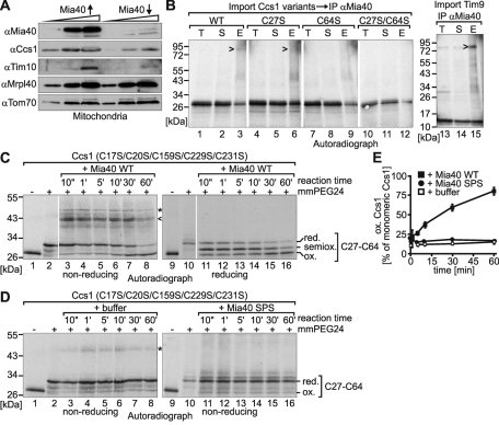
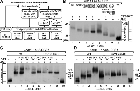
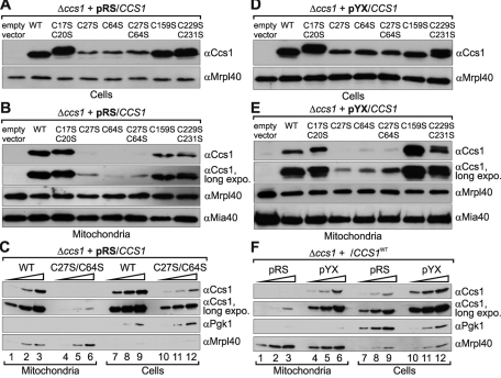
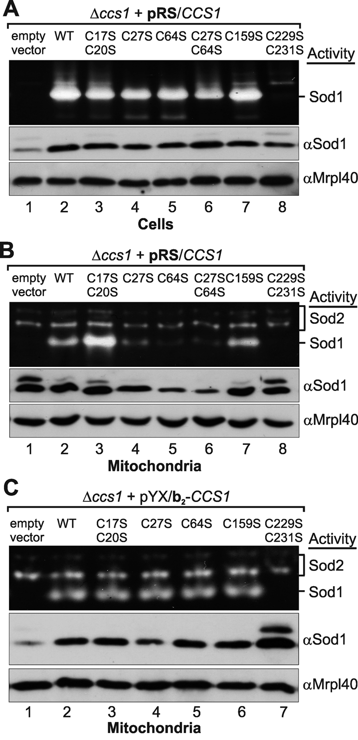
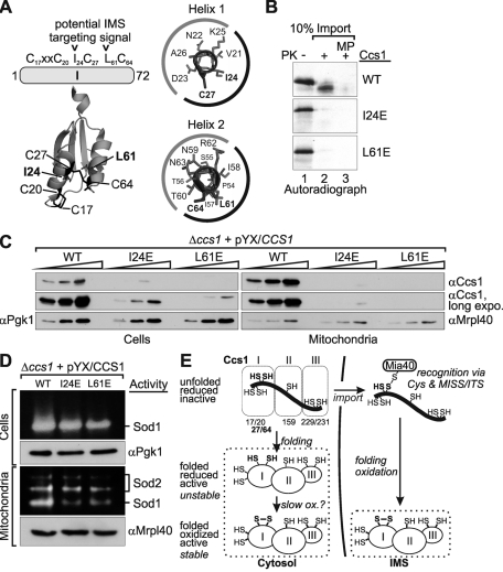
Superoxide dismutase 1 (Sod1) is an important antioxidative enzyme that converts superoxide anions to hydrogen peroxide and water. Active Sod1 is a homodimer containing one zinc ion, one copper ion, and one disulfide bond per subunit. Maturation of Sod1 depends on its copper chaperone (Ccs1). Sod1 and Ccs1 are dually localized proteins that reside in the cytosol and in the intermembrane space of mitochondria. The import of Ccs1 into mitochondria depends on the mitochondrial disulfide relay system. However, the exact mechanism of this import process has been unclear. In this study we detail the import and folding pathway of Ccs1 and characterize its interaction with the oxidoreductase of the mitochondrial disulfide relay Mia40. We identify cysteines at positions 27 and 64 in domain I of Ccs1 as critical for mitochondrial import and interaction with Mia40. On interaction with Mia40, these cysteines form a structural disulfide bond that stabilizes the overall fold of domain I. Although the cysteines are essential for the accumulation of functional Ccs1 in mitochondria, they are dispensable for the enzymatic activity of cytosolic Ccs1. We propose a model in which the Mia40-mediated oxidative folding of domain I controls the cellular distribution of Ccs1 and, consequently, active Sod1.


Figures

FIGURE 1:
FIGURE 1:
Cys-27 and Cys-64 are required for import of Ccs1 into isolated mitochondria. (A) Domain organization of yeast Ccs1. The three domains and the sequence positions of the seven cysteines of Ccs1 are depicted. (B) Import of Ccs1 into isolated mitochondria. In vitro translated radioactive Ccs1 (lanes 1 and 6, 10% control) was incubated with 50 μg of mitochondria isolated from Mia40-overexpressing strains. Subsequently, mitochondria were either analyzed directly (lane 2), treated with 100 μg/μl PK to remove nonimported proteins (lane 3), or turned into mitoplasts (MP) by hypotonic swelling and then treated with PK (lane 4) to remove all IMS proteins. One sample was also completely lysed with Triton X-100 (TX) and incubated with PK to test Ccs1 for protease resistance (lane 5). All samples were analyzed by reducing SDS–PAGE and autoradiography. The treatments with PK on mitochondria and mitoplasts were verified by immunoblotting against Erv1, Tim10 (both IMS), and Mrpl40 (matrix). (C) Glutathione dependence of Ccs1 import. Same as B, except that in vitro translated Ccs1 was incubated with different amounts of reduced glutathione during import. Experiments were quantified with ImageJ. (D) Import of Ccs1 cysteine mutants into isolated mitochondria. Same as B, except that in vitro translated Ccs1 cysteine mutants were incubated with mitochondria. Only whole mitochondria and mitoplasts were analyzed after PK digest. (E) Structure of domain I of Ccs1 (Lamb et al., 1999). This domain encompasses residues 2–74 and contains four cysteines (C17, C20, C27, and C64). Note that the C27–C64 disulfide connects two antiparallel α-helices, resulting in a structure of domain I that is similar to the structure of twin CX3C or twin CX9C substrates. (F) Import of Ccs1–domain I cysteine mutants and DHFR fusion variants into isolated mitochondria. Same as D, except that in vitro translated variants of domain I– or domain I–DHFR fusion proteins were incubated with mitochondria.
FIGURE 2:
FIGURE 2:
A disulfide bond between Cys-27 and Cys-64 of Ccs1 can be formed by Mia40 in vitro. (A) Protein levels in mitochondria isolated from strains with varying Mia40 levels. The yeast strain GalL–Mia40 was used to regulate the protein levels of Mia40. To overexpress Mia40 (Mia40↑), cells were grown in the presence of galactose. To deplete Mia40 (Mia40↓), cells were cultured in lactate medium containing 0.1% glucose for 16 h. Subsequently, mitochondria were isolated, and 10, 30, and 60 μg of isolated mitochondria were analyzed by SDS–PAGE and Western blotting with the indicated antibodies. (B) Interaction of Ccs1 variants with Mia40 during import into isolated mitochondria. Radiolabeled Ccs1 variants or the twin CX3C protein Tim9 were incubated with mitochondria isolated from a strain overexpressing Mia40 for 8 min at 25°C. Thiol–disulfide exchange reactions were stopped by addition of 50 mM NEM, mitochondria were lysed under denaturing conditions and diluted with Triton X-100–containing buffer, and supernatants were subjected to immunoprecipitation (IP) against Mia40. Bound material was analyzed by nonreducing SDS–PAGE and autoradiography. E, elution from beads after IP; T, total supernatant after IP. (C) In vitro oxidation of full-length Ccs1C17S/C20S/C159S/C229S/C231S by Mia40-WT. The in vitro translated radioactive Ccs1 mutant was incubated with 30 μM purified wild-type Mia40 (Mia40-WT) for the indicated times. Subsequently, samples were TCA precipitated, treated with 10 mM mm-PEG24 for 1 h, subjected to nonreducing (lanes 1–8) or reducing (lanes 9–16) SDS–PAGE, and analyzed by autoradiography. Arrowhead, disulfide-linked complex of Mia40 and Ccs1; asterisk, background band—potentially a disulfide-linked Ccs1 dimer. (D) As in C, except that the in vitro translated radioactive Ccs1 mutant was incubated with buffer (lanes 3–8) or 30 μM purified redox-inactive Mia40-SPS (lanes 11–16) for the indicated times. The samples were subjected to nonreducing SDS–PAGE. Asterisk, background band—potentially a disulfide-linked Ccs1 dimer. (E) Quantification of C and D. The autoradiographs of three different experiments were quantified using ImageJ.
FIGURE 3:
FIGURE 3:
In vivo Cys-27 and Cys-64 form a structural disulfide bond in IMS-localized Ccs1. (A) Scheme depicting the setup of the in vivo redox state measurements. (B) Migration standard for AMS alkylation experiments. Cells expressing different cysteine-to-serine mutants of Ccs1 were lysed with Triton X-100 (TX) and incubated with 20 mM DTT at 96°C for 10 min. Subsequently, proteins were precipitated with TCA and modified with 15 mM AMS, except for the sample in lane 8, which was left unmodified. This protocol ensures complete reduction of all cysteines and thus their complete AMS modification. The number of AMS moieties added to the proteins is indicated. The samples were analyzed by Western blotting against Ccs1. (C) In vivo redox state of wild-type (WT) Ccs1 and Ccs1C27S/C64S expressed under the control of the endogenous promoter of Ccs1 from a cen plasmid (pRS). Cells were treated as indicated according to the protocol described in A. Redox states at steady state are depicted in lanes 2 (WT) and 9 (Ccs1C27S/C64S). Note that wild-type Ccs1 can be fully reduced (i.e., modified with seven AMS) only upon boiling with 20 mM DTT at 96°C. (D) As in C, except that the in vivo redox state of overexpressed mitochondrial Ccs1 (pYX/b2CCS1) was determined.
FIGURE 4:
FIGURE 4:
Mutations of Cys-27 and Cys-64 result in decreased mitochondrial levels of Ccs1. (A) Ccs1 protein levels in Δccs1 cells expressing different Ccs1 variants under the control of the endogenous promoter (pRS). Cells were grown to mid-log phase in S(Lac)-Trp medium, lysed, and analyzed by SDS–PAGE and Western blotting against the indicated proteins. (B) Ccs1 protein levels in mitochondria isolated from Δccs1 cells expressing different Ccs1 variants under the control of the endogenous promoter from a cen plasmid (pRS). Cells were grown to mid-log phase in S(Lac)-Trp medium and lysed, and mitochondria were isolated. To remove proteins attached to the outside of mitochondria, the mitochondrial fraction was treated with 100 μg/ml PK. Mitochondrial fractions were analyzed by SDS–PAGE and immunoblotting using antibodies directed against Ccs1, Mrpl40 (mitochondrial matrix), and Mia40 (IMS). (C) Comparison of the cellular distribution of wild-type Ccs1 and Ccs1C27S/C64S in cells with endogenous levels of Ccs1. Experiments were performed as in A and B. Amounts of 5, 10, and 20 μg of mitochondria and cells at OD600 values of 0.05, 0.1, and 0.2 were analyzed. (D) Ccs1 protein levels in Δccs1 cells expressing different Ccs1 variants under the control of the triosephosphate isomerase promoter from a 2μ plasmid (pYX). As in A, except that Ccs1 was overexpressed in Δccs1 cells. (E) Ccs1 protein levels in mitochondria isolated from Δccs1 cells expressing different Ccs1 variants under the control of the triosephosphate isomerase promoter (pYX). As in B, except that Ccs1 was overexpressed in Δccs1 cells. (F) Comparison of the cellular distribution of wild-type Ccs1 in cells with endogenous levels of Ccs1 and cells overexpressing Ccs1. Experiments were performed as in A and B on Δccs1 cells expressing wild-type Ccs1 under the control of either the endogenous promoter (pRS) or the triosephosphate isomerase promoter (pYX). Totals of 5, 10, and 20 μg mitochondria and cells at OD600 values of 0.05, 0.1, and 0.2 were analyzed.
FIGURE 5:
FIGURE 5:
Levels of active mitochondrial Sod1 are lower in strains expressing the C27S and C64S cysteine mutants of Ccs1. (A) Sod1 protein and activity levels in Δccs1 cells expressing different Ccs1 variants under the control of the endogenous promoter (pRS). Cells were grown to mid-log phase in S(Lac)-Trp medium. Subsequently, cells were either lysed in native lysis buffer and analyzed by Sod activity gels (top) or lysed in SDS lysis buffer and analyzed by SDS–PAGE and Western blotting against the indicated proteins (middle and bottom). (B) Sod1 protein and activity levels in mitochondria isolated from Δccs1 cells expressing different Ccs1 variants under the control of the endogenous promoter from a cen plasmid (pRS). Cells were grown to mid-log phase in S(Lac)-Trp medium and lysed, and mitochondria were isolated. To remove proteins attached to the outside of mitochondria, the mitochondrial fraction was treated with 100 μg/ml PK. Mitochondrial fractions were then analyzed by Sod activity gels or by SDS–PAGE and immunoblotting using antibodies directed against Sod1, Mrpl40 (mitochondrial matrix), and Mia40 (IMS). (C) Sod1 activity and protein level in mitochondria isolated from cells expressing different b2–Ccs1 cysteine mutants. As in B, except that Ccs1 variants were fused to a b2-targeting sequence and expressed under the control of the triosephosphate isomerase promoter from a 2μ plasmid (pYX).
FIGURE 6:
FIGURE 6:
MISS/ITS mutants in Ccs1 behave like the C27S and C64S mutants of Ccs1. (A) Scheme of domain I of Ccs1 and location of the amino acids forming the putative MISS/ITS. Locations of amino acids forming the potential MISS/ITS are indicated in the structure of Ccs1 domain I, as well as in the helical-wheel representation of the two helices forming domain I. The black and gray labels of the wheel indicate the hydrophobic and hydrophilic faces of the helices, respectively. (B) Import of MISS/ITS mutants of Ccs1 into isolated mitochondria. The experiment was performed as indicated in Figure 1D. (C) Cellular distribution of the MISS/ITS mutants of Ccs1. The experiment was performed as indicated in Figure 4. (D) Sod activity in cells and mitochondria from CCS1 deletion strains expressing MISS/ITS mutants of Ccs1. The experiment was performed as indicated in Figure 5. (E) Model for the mechanism of Ccs1 distribution between the cytosol and the IMS. See Discussion for details.

Similar articles

Cited by

References

    1. Appenzeller-Herzog C, Ellgaard L. In vivo reduction-oxidation state of protein disulfide isomerase: the two active sites independently occur in the reduced and oxidized forms. Antioxid Redox Signal. 2008;10:55–64. - PubMed
    1. Appenzeller-Herzog C, Riemer J, Christensen B, Sorensen ES, Ellgaard L. A novel disulphide switch mechanism in Ero1alpha balances ER oxidation in human cells. EMBO J. 2008;27:2977–2987. - PMC - PubMed
    1. Banci L, et al. Molecular chaperone function of Mia40 triggers consecutive induced folding steps of the substrate in mitochondrial protein import. Proc Natl Acad Sci USA. 2010;107:20190–20195. - PMC - PubMed
    1. Bien M, Longen S, Wagener N, Chwalla I, Herrmann JM, Riemer J. Mitochondrial disulfide bond formation is driven by intersubunit electron transfer in Erv1 and proofread by glutathione. Mol Cell. 2010;37:516–528. - PubMed
    1. Chacinska A, Koehler CM, Milenkovic D, Lithgow T, Pfanner N. Importing mitochondrial proteins: machineries and mechanisms. Cell. 2009;138:628–644. - PMC - PubMed

Publication types

MeSH terms

Substances