. 2011 Oct;22(20):3840-52.
doi: 10.1091/mbc.E11-04-0364. Epub 2011 Aug 24.

ARHGAP18, a GTPase-activating protein for RhoA, controls cell shape, spreading, and motility

Affiliations

ARHGAP18, a GTPase-activating protein for RhoA, controls cell shape, spreading, and motility

Masao Maeda et al. Mol Biol Cell. 2011 Oct.

Abstract

Rho GTPases are molecular switches that transmit biochemical signals in response to extracellular stimuli to elicit changes in the actin cytoskeleton. Rho GTPases cycle between an active, GTP-bound state and an inactive, GDP-bound state. These states are regulated by two distinct families of proteins-guanine nucleotide exchange factors and GTPase-activating proteins (GAPs). We studied the role of a previously uncharacterized GAP, ARHGAP18 (MacGAP). Overexpression of ARHGAP18 suppressed the activity of RhoA and disrupted stress fiber formation. Conversely, silencing of ARHGAP18 by small interfering RNA transfection-enhanced stress fiber formation and induced rounding of cells. We examined the role of ARHGAP18 in cell spreading and migration. Immunofluorescence analysis revealed that ARHGAP18 was localized to the leading edge during cell spreading and migration. ARHGAP18-knockdown cells showed impaired spreading, premature formation of stress fibers, and sustained activation of RhoA upon cell attachment. In addition, knockdown and overexpression of ARHGAP18 resulted in the inhibition and promotion of cell migration, respectively. Furthermore, ARHGAP18 was required for the polarization of cells for migration. Our results define ARHGAP18 as one of the crucial factors for the regulation of RhoA for the control of cell shape, spreading, and migration.


Figures
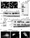
FIGURE 1:
FIGURE 1:
Expression and localization of ARHGAP18 in cells. (A) HeLa cells were cultured in 24-well plates and transfected with Luc or ARHGAP18 siRNA. Seventy-two hours later, cells were fixed and stained with FITC-labeled paclitaxel to visualize cells. Scale bar, 50 μm. (B) HeLa cells were transfected with indicated siRNAs, and 72 h later, the cells were lysed and immunoblotted with anti-ARHGAP18 antibody. (C) Expression of ARHGAP18 in cell lines from various tissues. (D) NIH3T3 cells were transfected with either Luc or mouse ARHGAP18 siRNA, and 72 h later, cells were lysed and the expression was examined by immunoblot. (E) Expression of ARHGAP18 and RhoA in mouse tissues was examined by immunoblot. (F) HeLa cells transfected with Luc or ARHGAP18 siRNA were fixed and immunostained with anti-ARHGAP18 antibody. Scale bar, 20 μm. (E) GFP-tagged ARHGAP18 was transiently expressed in HeLa cells. Scale bar, 20 μm.
FIGURE 2:
FIGURE 2:
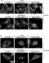
Suppression of ARHGAP18 affects stress fiber and focal adhesion formation. (A) HeLa and MDA-MB-231 cells cultured on the fibronectin-coated glass coverslips were transfected with Luc or ARHGAP18 siRNA and 3 d later, cells were fixed and stained with rhodamine-conjugated phalloidin. Scale bar, 20 μm. (B) HeLa and MDA-MB-231 cells were treated as in A and immunostained with anti-vinculin antibody. Scale bar, 20 μm.
FIGURE 3:
FIGURE 3:
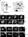
Overexpression of wild-type ARHGAP18, but not GAP-defective ARHGAP18, suppresses formation of stress fibers and focal adhesions. (A) Expression of ARHGAP18 in each cell line was examined with anti-ARHGAP18 and anti-GFP antibodies. FL, full-length ARHGAP18; ΔGAP, ARHGAP18 with the deletion of RhoGAP domain; R365A, full-length ARHGAP18 with the substitution of arginine at 365 to alanine. Arrows indicate endogenous ARHGAP18. (B) Cells cultured on the fibronectin-coated glass coverslips were fixed and stained with rhodamine-conjugated phalloidin. Scale bar, 20 μm. (C) Cells cultured on the fibronectin-coated glass coverslips were fixed and stained with anti-vinculin antibody. Scale bar, 20 μm.
FIGURE 4:
FIGURE 4:
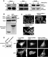
Regulation of RhoA activity by ARHGAP18 controls stress fiber formation. (A) HeLa cells that constitutively expressed either GFP or GFP-tagged ARHGAP18 were lysed and mixed with GST-Rhotekin-RBD (RhoA) or GST-PAK-PBD (Rac1 and Cdc42) bound to glutathione-agarose beads to precipitate the active form of Rho GTPases. The immunoprecipitates were subjected to immunoblot analysis with the indicated antibodies. (B) HeLa cells that constitutively expressed GFP-tagged ARHGAP18 were lysed and mixed with either GST or GST-RhoA (Q63L) coupled to glutathione-agarose beads, and interacting proteins were affinity precipitated. The immunoprecipitates were subjected to immunoblot analysis with anti-GFP antibody to determine the association of GFP-tagged ARHGAP18 and RhoA (Q63L). Bottom, Coomassie blue staining of recombinant proteins. (C) HeLa cells cultured on the fibronectin-coated glass coverslips were transfected with the indicated siRNAs. Three days later, ARHGAP18 siRNA–transfected cells were treated with DMSO or Y27632 for 1 h and immunostained with rhodamine-conjugated phalloidin. Scale bar, 20 μm. (D) HeLa cells were infected with recombinant virus that encoded the shRNA targeting either luciferase (Luc) or ARHGAP18 and selected with puromycin. Expression of ARHGAP18 in each cell line was examined by immunoblot analysis. Cells were transfected with GFP or GFP-tagged dominant-negative RhoA (T19N) and 48 h later, cells were fixed and immunostained with rhodamine-conjugated phalloidin. Scale bar, 20 μm.
FIGURE 5:
FIGURE 5:
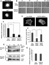
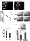
Inactivation of RhoA by ARHGAP18 is necessary for prompt cell spreading. (A) HeLa cells were seeded on fibronectin-coated surfaces and fixed 1 h later. Cells were immunostained with anti-ARHGAP18 or anti-GST antibody. Scale bar, 20 μm. (B) HeLa cells were transfected with either Luc or ARHGAP18 siRNA, and 72 h later, suspended cells were seeded onto fibronectin-coated dishes. Cell spreading was monitored by time-lapse microscopy. Representative images are shown. Scale bar, 20 μm. (C) siRNA-transfected HeLa cells were seeded on the fibronectin-coated dishes, and 1 h later, cells were fixed and evaluated for the ratio of spread cells. The graph shows ratios of spread cells counted from five random fields of three independent experiments (means ± SD; *p < 0.01). (D) siRNA-transfected HeLa cells were plated on fibronectin-coated glass coverslips, and 1 h later, cells were fixed and immunostained with rhodamine-conjugated phalloidin. scale bar, 20 μm. (E) HeLa/shLuc or HeLa/shGAP18 cells were seeded on fibronectin-coated dishes and lysed at the indicated time points. Cell lysates were mixed with GST-Rhotekin-RBD coupled to glutathione-agarose beads to precipitate active RhoA. The immunoprecipitates were subjected to immunoblot analysis with anti-RhoA antibody. (F) siRNA-transfected HeLa cells were seeded on the fibronectin-coated surface, and 15 min later either DMSO or Y27632 was added. One hour after seeding, the cells were fixed and counted for the ratio of spread cells. The graphs indicate the ratio of spread cells counted in five randomly selected fields from three independent experiments (means ± SD; *p < 0.01). (G) Cell attachment assays of Luc and GAP18 siRNA–transfected HeLa cells. Cells (1 × 10⁵) were seeded onto fibronectin-coated 24-well plates, and 20 min later, unattached cells were washed out and attached cells in five randomly selected fields were counted. The graph shows the relative ratio of attached cells from three independent experiments (means ± SD; *p < 0.01).
FIGURE 6:
FIGURE 6:
ARHGAP18 regulates cell migration. (A) Confluent monolayers of MDA-MB-231 cells were scratched and fixed 4 h later. Cells were immunostained with anti-ARHGAP or anti-GST antibody. (B) MDA-MB-231 cells were transfected with Luc or ARHGAP18 siRNA, and 3 d later, a scratch was made and migration was examined for 18 h. The graph shows the distance of the wound measured in five randomly selected points from three independent experiments (means ± SD; *p < 0.01). Right, representative images of cells in the assay. Scale bar, 500 μm. (C) siRNA-transfected MDA-MB-231 cells were loaded onto the upper surface of Boyden chambers, incubated for 4 h, fixed, and examined by microscopy. The graph indicates the relative ratio of cells that migrated to the lower surface of the filter from five randomly selected fields in three independent experiments (means ± SD; *p < 0.01). (D) MDA-MB-231 cells that constitutively expressed GFP or GFP-tagged ARHGAP18 were established by infecting with recombinant retrovirus. Expression of ARHGAP18 was examined by immunoblot. The arrow indicates endogenous ARHGAP18 and the arrowhead GFP-ARHGAP18. Migration of these cells was examined using Boyden chambers. The graph indicates the relative ratio of cells that migrated to the lower surface of the filter from five randomly selected fields in three independent experiments (means ± SD; *p < 0.01).
FIGURE 7:
FIGURE 7:
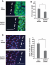
ARHGAP18 controls cellular polarity for migration. (A) A confluent monolayer of KMST-6 cells transfected with each siRNA was scratched and incubated for 4 h. Cells were fixed and immunostained with anti–α-tubulin antibody and DAPI. White lines indicate wound direction. Scale bar, 100 μm. (B) The length of protrusions of cells on the wound edge treated as in A was measured. Fifty cells from randomly selected fields were measured in each of three independent experiments. Data are shown as average distances (mean ± SD) between the leading edge and the nucleus (*p < 0.01). (C) siRNA-transfected KMST-6 cells were wounded and incubated for 4 h. Cells were fixed and immunostained with anti-GM130 antibody and DAPI to evaluate the percentage of cells with Golgi located in the 120° arc facing the wound. One hundred cells on the wound edge were evaluated for Golgi localization in each of three independent experiments (mean ± SD; *p < 0.01). Left, representative images of immunostained cells 4 h after wounding. White lines indicate wound direction. Red, GM130; blue, DAPI. Scale bar, 100 μm.

Similar articles

Cited by

References

    1. Amano M, Nakayama M, Kaibuchi K. Rho-kinase/ROCK: a key regulator of the cytoskeleton and cell polarity. Cytoskeleton (Hoboken) 2010;67:545–554. - PMC - PubMed
    1. Arthur WT, Burridge K. RhoA inactivation by p190RhoGAP regulates cell spreading and migration by promoting membrane protrusion and polarity. Mol Biol Cell. 2001;12:2711–2720. - PMC - PubMed
    1. Arthur WT, Noren NK, Burridge K. Regulation of Rho family GTPases by cell-cell and cell-matrix adhesion. Biol Res. 2002;35:239–246. - PubMed
    1. Arthur WT, Petch LA, Burridge K. Integrin engagement suppresses RhoA activity via a c-Src-dependent mechanism. Curr Biol. 2000;10:719–722. - PubMed
    1. Barrett T, Xiao B, Dodson EJ, Dodson G, Ludbrook SB, Nurmahomed K, Gamblin SJ, Musacchio A, Smerdon SJ, Eccleston JF. The structure of the GTPase-activating domain from p50rhoGAP. Nature. 1997;385:458–461. - PubMed

Publication types

MeSH terms