. 2011 Oct;22(20):3812-25.
doi: 10.1091/mbc.E11-04-0328. Epub 2011 Aug 24.

Serum- and glucocorticoid-induced kinase 3 in recycling endosomes mediates acute activation of Na+/H+ exchanger NHE3 by glucocorticoids

Affiliations

Serum- and glucocorticoid-induced kinase 3 in recycling endosomes mediates acute activation of Na+/H+ exchanger NHE3 by glucocorticoids

Peijian He et al. Mol Biol Cell. 2011 Oct.

Abstract

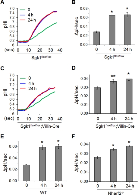
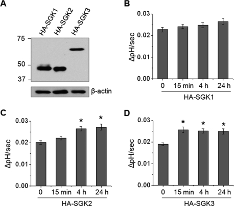
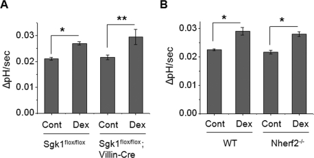
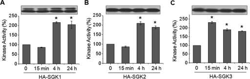
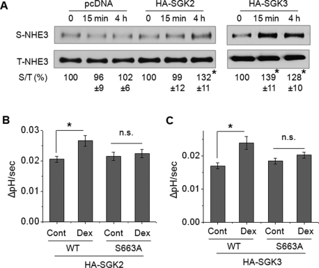
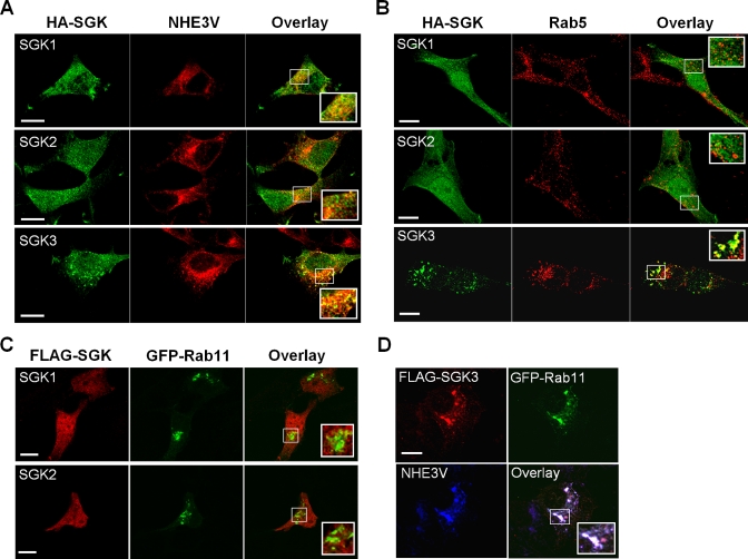
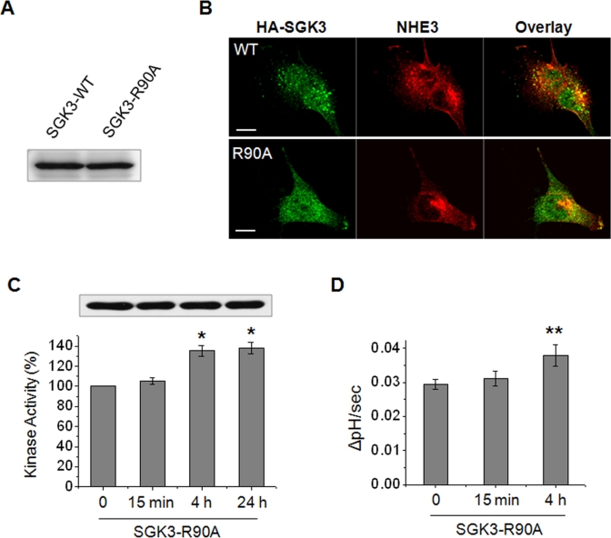
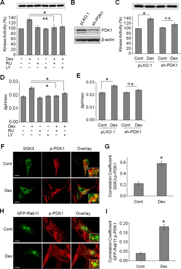
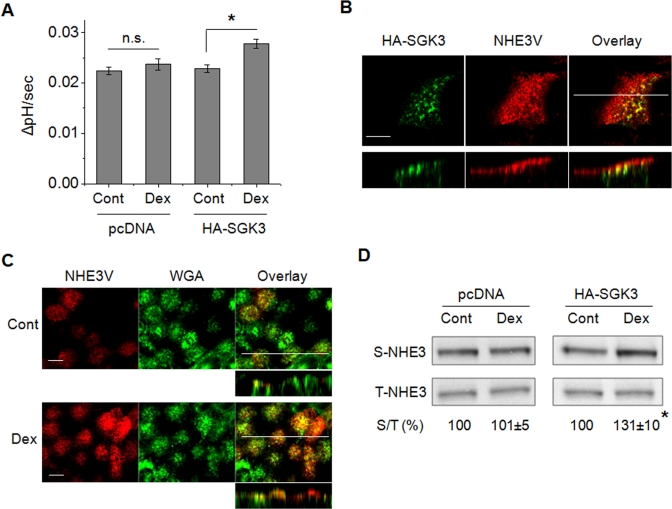
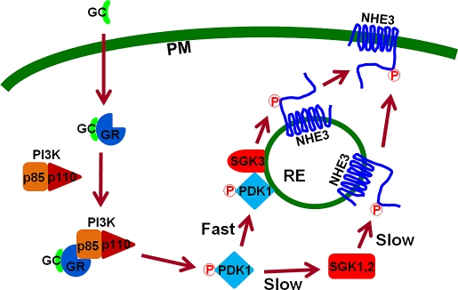
Na(+)/H(+) exchanger 3 (NHE3) is the major Na(+) transporter in the intestine. Serum- and glucocorticoid-induced kinase (SGK) 1 interacts with NHE regulatory factor 2 (NHERF2) and mediates activation of NHE3 by dexamethasone (Dex) in cultured epithelial cells. In this study, we compared short-term regulation of NHE3 by Dex in SGK1-null and NHERF2-null mice. In comparison to wild-type mice, loss of SGK1 or NHERF2 significantly attenuated regulation of NHE3 by Dex but did not completely obliterate the effect. We show that transfection of SGK2 or SGK3 in PS120 cells resulted in robust activation of NHE3 by Dex. However, unlike SGK1 or SGK2, SGK3 rapidly activated NHE3 within 15 min of Dex treatment in both PS120 and Caco-2bbe cells. Immunofluorescence analysis showed that SGK3 colocalized with NHE3 in recycling endosomes, whereas SGK1 and SGK2 were diffusely distributed. Mutation of Arg-90 of SGK3 disrupted the endosomal localization of SGK3 and delayed NHE3 activation. Activation of SGK3 and NHE3 by Dex was dependent on phosphoinositide 3-kinase (PI3K) and phosphoinositide-dependent kinase 1 (PDK1), and Dex induced translocation of PDK1 to endosomes. Our study identifies SGK3 as a novel endosomal kinase that acutely regulates NHE3 in a PI3K-dependent mechanism.


Figures

FIGURE 1:
FIGURE 1:
SGK1 and NHERF2 play important roles in Dex-induced activation of NHE3 in mouse ileum. Mice were injected intraperitoneally with Dex at 2 mg/kg body weight. After 4 or 24 h, villi were isolated from the ileum and NHE3 activity was measured in the presence of 50 μM Hoe-694 (NHE1 and NHE2 inhibitor). Increased rates of pH recovery following 4 or 24 h of Dex treatment were observed. (A) Representative traces of Na+-dependent pH recovery in Sgk1flox/flox mice are shown. (B) NHE3 activities in Sgk1flox/flox villi are presented as the rate of Na+-dependent pH change by determining ΔpHi/Δtime at pHi 6.5 using typical traces shown in (A). NHE3 activities are shown as mean ± SE. (C) Representative traces of Na+-dependent pH recovery in the ileal villi prepared from Sgk1flox/flox;Villin-Cre mice are shown. (D) NHE3 activities in Sgk1flox/flox;Villin-Cre villi are presented as the rate of Na+-dependent pH change at pHi 6.5. Stimulation of NHE3-dependent pH recovery is significantly attenuated in Sgk1flox/flox;Villin-Cre mice compared with Sgk1flox/flox mice. NHE3 activities at pHi 6.5 are shown in (E) WT and (F) Nherf2−/− mice. For all experiments, n = 3–4 mice and 6–8 villi per mouse were used. *, p < 0.01 and **, p < 0.05, compared with the untreated control.
FIGURE 2:
FIGURE 2:
SGK3 rapidly activates NHE3 in response to Dex. (A) PS120/NHE3V cells were stably transfected with HA-SGK1, HA-SGK2, and HA-SGK3, and the expression levels of SGKs were determined by immunoblotting using an anti-HA antibody. Western blot of β-actin was used as loading control. PS120/NHE3V cells expressing (B) HA-SGK1, (C) HA-SGK2, or (D) HA-SGK3 were treated with 1 μM Dex for t = 0, 15 min, 4 h, or 24 h, followed by determination of NHE3 activity. The rates of pH recovery at pHi 6.7 are shown. n = 8. *, p < 0.01 compared with the untreated control.
FIGURE 3:
FIGURE 3:
Dex acutely activates NHE3 in mouse intestine lacking SGK1 or NHERF2. (A) Villi were isolated from the Sgk1flox/flox and Sgk1flox/flox;Villin-Cre ilea and were treated ex vivo with 1 μM Dex or carrier for 15 min prior to measurement of NHE3 activity in the presence of Hoe-694. The rates of pH recovery at pHi 6.6 are shown. (B) The rates of pH recovery at pHi 6.6 in WT and Nherf2−/− ilea treated with Dex for 15 min are shown. For all experiments, n = 3 mice and six villi per mouse were used. *, p < 0.01 and **, p < 0.05, compared with the control.
FIGURE 4:
FIGURE 4:
SGK3 kinase activity is acutely stimulated by Dex. PS120/NHE3V cells expressing HA-SGK1, HA-SGK2, or HA-SGK3 were treated with 1 μM Dex for t = 0, 15 min, 4 h, or 24 h. SGK proteins were purified by immunoprecipitation with an anti-HA antibody. To ensure the equal loading of immunoprecipitated SGKs, an aliquot of eluted protein was subjected to Western blotting using anti-HA antibody. Equal amounts of each SGK protein were used for in vitro kinase assay by using [γ-32P]ATP, as detailed in Materials and Methods. The activities of (A) SGK1, (B) SGK2, and (C) SGK3 are shown relative to the basal SGK activity. n = 3. *, p < 0.01 compared with the untreated control.
FIGURE 5:
FIGURE 5:
SGK2 and SGK3 increase NHE3 surface expression and the S663A mutation blocks the activation. (A) PS120/NHE3V/pcDNA, PS120/NHE3V/HA-SGK2, and PS120/NHE3V/HA-SGK3 cells were treated with 1 μM Dex for t = 0, 15 min, or 4 h, and the amount of NHE3 protein on the plasma membrane was determined by surface biotinylation, as detailed in Materials and Methods. The amount of surface (S) NHE3 was normalized to total (T) NHE3, and relative changes (%) are shown below the Western blots. n ≥ 3. (B) PS120/OKNHE3/HA-SGK2 and PS120/OKNHE3-S663A/HA-SGK2 cells were treated with 1 μM Dex for 4 h and NHE3 activity was measured. The rates of pH recovery at pHi 6.8 are shown. n = 6. (C) PS120/OKNHE3/HA-SGK3 and PS120/OKNHE3-S663A/HA-SGK3 cells were treated with 1 μM Dex for 15 min, and NHE3 activity was determined. The rates of pH recovery at pHi 6.8 are shown. n = 6. n.s., not significant. *, p < 0.01, compared with the untreated control.
FIGURE 6:
FIGURE 6:
SGK3 colocalizes with NHE3 in recycling endosomes. (A) Cellular distribution of HA-tagged SGK1-3 (green) and NHE3V (red) in PS120 cells was determined by indirect immunofluorescence with anti-HA and anti-VSVG antibodies, respectively. The inset shows a magnified image of the boxed area. (B) Cellular localization of HA-tagged SGK1-3 (green) and Rab5 (red) in PS120 cells was determined with anti-HA and anti-Rab5 antibodies, respectively. (C) PS120/NHE3V cells were transfected with FLAG-tagged SGK1 or SGK2 and GFP-Rab11. The colocalization of SGK1 or SGK2 with Rab11 was determined with anti-FLAG antibody. (D) PS120/NHE3V cells were transfected with FLAG-SGK3 and GFP-Rab11. The colocalization of SGK3 (red) and NHE3V (blue) with GFP-Rab11 (green) was determined with anti-FLAG and anti-VSVG antibodies, respectively. Scale bar: 10 μm.
FIGURE 7:
FIGURE 7:
Acute regulation of NHE3 by SGK3 is dependent on the endosomal localization of SGK3. (A) PS120/NHE3V cells were expressed with HA-SGK3 or HA-SGK3-R90A, and the expression levels of SGK3 and SGK3-R90A in PS120/NHE3V cells were examined with anti-HA antibody. (B) Cellular distribution of SGK3 (green) or SGK3-R90A (green) with NHE3 (red) in PS120 cells was examined with anti-HA and anti-VSVG antibodies, respectively. Scale bar: 10 μm. (C) PS120/NHE3V/HA-SGK3-R90A cells were treated with Dex for t = 0, 15 min, 4 h, or 24 h, and SGK3-R90A protein was purified by immunoprecipitation for kinase assay. The kinase activities of SGK3-R90A are shown relative to the basal activity. n = 3. (D) PS120/NHE3V/HA-SGK3-R90A cells were treated with 1 μM Dex for t = 0, 15 min, or 4 h, followed by measurement of NHE3 activity. The rates of pH recovery at pHi 6.7 are shown. n = 6. *, p < 0.01 and **, p < 0.05, compared with the untreated control.
FIGURE 8:
FIGURE 8:
SGK3-mediated activation of NHE3 is dependent on PI3K and PDK1. (A) PS120/NHE3V/HA-SGK3 cells were pretreated with 1 μM RU or 20 μM LY prior to treatment with 1 μM Dex or carrier for 15 min. SGK3 protein was purified by immunoprecipitation, and the kinase activity was determined by TR-FRET. n = 3. (B) PS120/NHE3V/HA-SGK3 cells were stably transfected with sh-PDK1 or pLKO.1, and the expression level of PDK1 was determined by Western blotting using an anti-PDK1 antibody. (C) PS120/NHE3V/HA-SGK3 cells transfected with sh-PDK1 or pLKO.1 were treated with 1 μM Dex or carrier for 15 min. SGK3 protein was purified, and kinase activity was determined. n = 3. (D) NHE3 activities were determined in PS120/NHE3V/HA-SGK3 cells pretreated with RU or LY prior to treatment with Dex. (E) NHE3 activities in PS120/NHE3V/HA-SGK3 cells transfected with sh-PDK1 or pLKO.1 are shown. (A–E) n.s., not significant. *, p < 0.01 and **, p < 0.05, compared with respective controls. n = 6. (F) PS120/NHE3V cells expressing HA-SGK3 were serum-starved and treated with Dex or carrier for 15 min. Colocalization of SGK3 (green) and phospho-PDK1(p-PDK1, red) was determined using anti-HA and anti-p-PDK1 antibodies, respectively. (G) The graph represents Pearson's coefficient of SGK3 and p-PDK1 colocalization from 10 independent fields of cells. *, p < 0.01 compared with the control. (H) Colocalization of GFP-Rab11 (green) and p-PDK1 (red) in PS120/NHE3V cells treated with or without Dex for 15 min is shown. Scale bar: 10 μm. Insets show high magnification of merged signals. (I) Pearson's coefficient of GFP-Rab11 and p-PDK1 colocalization from 10 independent fields of cells. *, p < 0.01 compared with the control.
FIGURE 9:
FIGURE 9:
SGK3 mediates an acute activation of NHE3 by Dex in Caco-2bbe cells. (A) Caco-2bbe/hNHE3V/HA-SGK3 and Caco-2bbe/hNHE3V/pcDNA cells were treated with Dex or carrier for 15 min. NHE3 activities were measured in the presence of 50 μM Hoe-694. The rates of pH recovery at pHi 6.7 are shown. n = 6. n.s., not significant. *, p < 0.01 compared with the untreated control. (B) Cellular localization of SGK3 (green) and NHE3 (red) in Caco-2bbe cells was determined by indirect immunofluorescence with anti-HA and anti-VSVG antibodies, respectively. Confocal focal planes (top) and cross-sections (bottom) are shown. Scale bar: 10 μm. (C) Caco-2bbe/hNHE3V/HA-SGK3 cells were treated with Dex or carrier for 15 min. NHE3 expression (red) at the apical membrane was determined with anti-VSVG antibody. WGA (green) staining of glycoprotein at plasma membrane is shown as a marker of apical labeling. Confocal focal (large panel) and cross-sectional (small panel) views are shown. Scale bar: 10 μm. (D) The amount of NHE3 protein on the plasma membrane was determined by surface biotinylation, as detailed in Materials and Methods. The amount of surface (S) NHE3 was normalized to total (T) NHE3, and relative changes (%) are shown below the blotting. n = 3. *, p < 0.01 compared with the untreated control.
FIGURE 10:
FIGURE 10:
A schematic model of GC-mediated acute activation of NHE3 via SGK3. Previous studies showed that GCs bind to GR, and activated GR interacts with the p85 regulatory subunit of PI3K and recruits the p110 catalytic subunit in the cytoplasm (Hafezi-Moghadam et al., 2002; Limbourg et al., 2002; Hu et al., 2009). PI3K activates PDK1 by phosphorylating PDK1, which acutely mobilizes to recycling endosomes (RE), where SGK3 and NHE3 are expressed. PDK1 then activates SGK3, which in turn phosphorylates NHE3 and induces exocytotic insertion of NHE3 into the plasma membrane (PM). This cascade occurs as part of the acute NHE3 activation by Dex involving SGK3. In the absence of SGK3, the activated PDK1 diffuses through cytoplasmic matrix and activates SGK1 or SGK2. Activated SGK1 and SGK2 then phosphorylate NHE3 at endosomes and potentiate trafficking of NHE3 to PM, a later pathway of NHE3 regulation.

Similar articles

Cited by

References

    1. Alvarez de la Rosa D, Zhang P, Naray-Fejes-Toth A, Fejes-Toth G, Canessa CM. The serum and glucocorticoid kinase sgk increases the abundance of epithelial sodium channels in the plasma membrane of Xenopus oocytes. J Biol Chem. 1999;274:37834–37839. - PubMed
    1. Baum M, Moe OW, Gentry DL, Alpern RJ. Effect of glucocorticoids on renal cortical NHE-3 and NHE-1 mRNA. Am J Physiol. 1994;267:F437–E442. - PubMed
    1. Biemesderfer D, Rutherford PA, Nagy T, Pizzonia JH, AbuAlfa AK, Aronson PS. Monoclonal antibodies for high-resolution localization of NHE3 in adult and neonatal rat kidney. Am J Physiol Renal Physiol. 1997;273:F289–F299. - PubMed
    1. Boehmer C, Henke G, Schniepp R, Palmada M, Rothstein JD, Broer S, Lang F. Regulation of the glutamate transporter EAAT1 by the ubiquitin ligase Nedd4-2 and the serum and glucocorticoid-inducible kinase isoforms SGK1/3 and protein kinase B. J Neurochem. 2003;86:1181–1188. - PubMed
    1. Bohmer C, Sopjani M, Klaus F, Lindner R, Laufer J, Jeyaraj S, Lang F, Palmada M. The serum and glucocorticoid inducible kinases SGK1-3 stimulate the neutral amino acid transporter SLC6A19. Cell Physiol Biochem. 2010;25:723–732. - PubMed

Publication types

MeSH terms