. 2011 Oct;22(20):3826-39.
doi: 10.1091/mbc.E11-06-0492. Epub 2011 Aug 24.

The functionally distinct fission yeast formins have specific actin-assembly properties

Affiliations

The functionally distinct fission yeast formins have specific actin-assembly properties

Bonnie J Scott et al. Mol Biol Cell. 2011 Oct.

Abstract

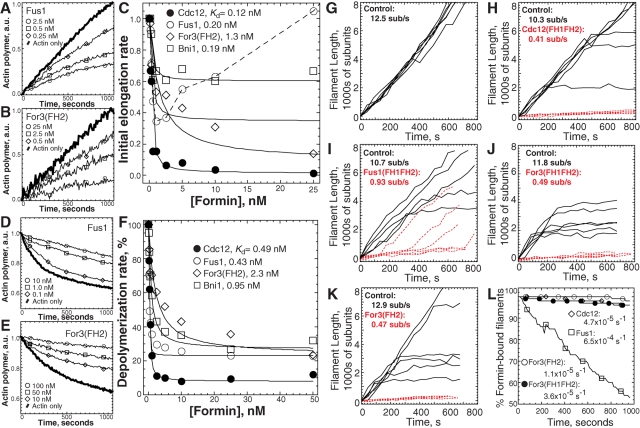
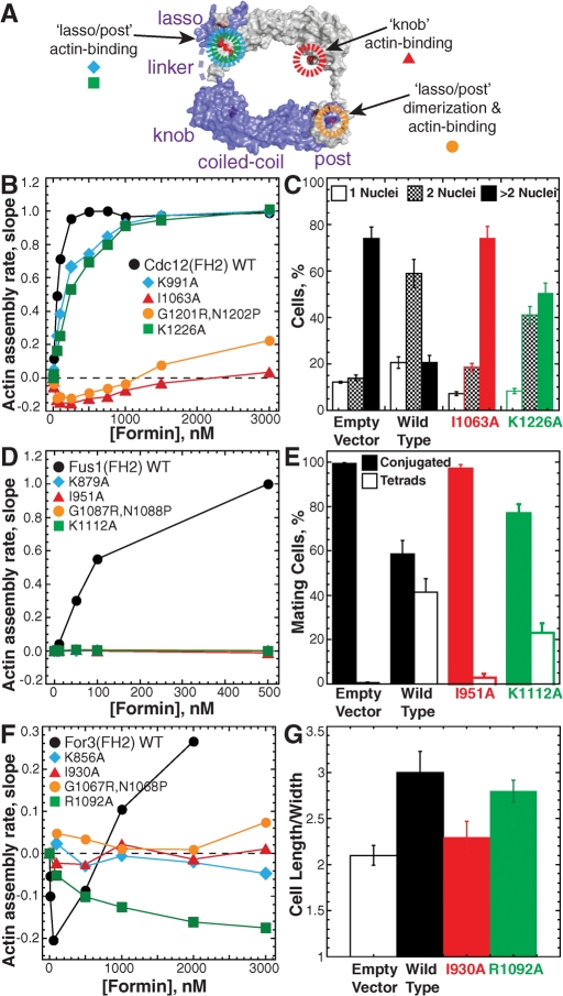
Fission yeast expresses three formins required for distinct actin cytoskeletal processes: Cdc12 (cytokinesis), For3 (polarization), and Fus1 (mating). We propose that in addition to differential regulation, key actin-assembly properties tailor formins for a particular role. In direct comparison to the well-studied Cdc12, we report the first in vitro characterization of the actin-assembly properties of For3 and Fus1. All three share fundamental formin activities; however, particular reaction rates vary significantly. Cdc12 is an efficient nucleator (one filament per approximately 3 Cdc12 dimers) that processively elongates profilin-actin at a moderate rate of 10 subunits s(-1) μM(-1), but lacks filament-bundling activity. Fus1 is also an efficient nucleator, yet processively elongates profilin-actin at one-half the rate of and dissociates 10-fold more rapidly than Cdc12; it also bundles filaments. For3 nucleates filaments 100-fold less well than Fus1, but like Cdc12, processively elongates profilin-actin at a moderate rate and lacks filament-bundling activity. Additionally, both the formin homology FH1 and FH2 domains contribute to the overall rate of profilin-actin elongation. We also confirmed the physiological importance of the actin-assembly activity of the fission yeast formins. Point mutants that disrupt their ability to stimulate actin assembly in vitro do not function properly in vivo.


Figures

FIGURE 1:
FIGURE 1:
The fission yeast formins stimulate actin filament assembly with different nucleation efficiencies. (A) Domain organization of the three fission yeast formins Cdc12, Fus1, and For3, and budding yeast formin Bni1. Amino acid residues delineating FH1 and FH2 domain constructs are shown to the right. Each “P” signifies a putative profilin binding site, which is a proline-rich segment of 6–14 residues. (B to F) Spontaneous assembly of 2.5 μM Mg-ATP actin monomers (20% pyrene-labeled). (B and C) Time course of actin assembly in the absence (thick curve) or presence of the indicated concentrations of Fus1(FH1FH2), For3(FH1FH2), and For3(FH2). (D) Normalized plot of the dependence of actin-assembly rate (slope) on concentration of Cdc12(FH1FH2) (□), Fus1(FH1FH2) (▵), For3(FH1FH2) (•), For3(FH2) (◯), and Bni1(FH1FH2) (⋄). The inset shows For3(FH2) (◯) with an expanded x-axis. Dashed lines indicate the zero coordinate on the y-axis. (E) Nucleation efficiency: optimal number of formin dimers required to nucleate one filament. (F) Representative fluorescence micrographs of rhodamine-phalloidin–labeled actin filaments after spontaneous actin-assembly reactions reached plateau (2 h) in the absence (actin only) or presence of the indicated formin. Average filament lengths are reported in μm ± SD. Scale bar: 10 μm.
FIGURE 2:
FIGURE 2:
The fission yeast formins bind to and reduce actin filament barbed-end dynamics by different amounts. (A to C) Seeded elongation: addition of 0.2 μM Mg-ATP-actin monomers (20% pyrene-labeled) to the barbed end of 0.5 μM preassembled actin filaments. (A and B) Time course of seeded assembly alone (thick curve) or in the presence of the indicated concentrations of Fus1(FH1FH2) (A) and For3(FH2) (B). (C) Dependence of the initial barbed-end assembly rate on formin concentration. Curve fits revealed equilibrium dissociation constants of 0.12 nM for Cdc12(FH1FH2) (•), 0.20 nM for Fus1(FH1FH2) (◯), 1.3 nM for For3(FH2) (⋄), and 0.19 nM for Bni1(FH1FH2) (□). Fus1(FH1FH2) concentrations ≥5.0 nM significantly nucleate actin monomer assembly under these conditions, so its curve fit was from 0 to 2.5 nM. (D to F) Filament disassembly: barbed-end loss of actin monomer from 5.0 μM preassembled filaments (50% pyrene labeled) upon dilution to 0.1 μM. (D-E) Depolymerization time-course in the absence (thick curve) or presence of the indicated concentrations of Fus1(FH1FH2) (D) and For3(FH2) (E). (F) Dependence of the depolymerization rate on formin concentration. Curve fits revealed equilibrium dissociation constants of 0.49 nM for Cdc12(FH1FH2) (•), 0.43 nM for Fus1(FH1FH2) (◯), 2.3 nM for For3(FH2) ⋄), and 0.95 nM for Bni1(FH1FH2) (□). (G to L) TIRF microscopy visualization of the spontaneous assembly of unlabeled 1.0 μM Mg-ATP-actin with 0.5 μM Mg-ATP-actin labeled with Oregon green. (G to K) Plots of the length of six individual filaments over time for control (solid lines) and formin-nucleated (red dashed lines) filaments. The average elongation rates are indicated (subunits s−1). (G) Actin only control (Video S1). (H) 1.0 nM Cdc12(FH1FH2) (Video S2). (I) 0.5 nM Fus1(FH1FH2) (Video S3). (J) 150 nM For3(FH1FH2) (Video S4). (K) 50 nM For3(FH2) (Video S5). (L) Dissociation rate. Percentage of formin-associated filaments over time. Curve fits revealed dissociation rates of 4.7 × 10⁻⁵ s−1 for Cdc12(FH1FH2) (⋄), 6.5 × 10⁻⁴ s−1 for Fus1(FH1FH2) (□), 3.6 × 10⁻⁵ s−1 for For3(FH1FH2) (•), and 1.1 × 10⁻⁵ s−1 for For3(FH2) (◯).
FIGURE 3:
FIGURE 3:
The fission yeast formins stimulate the assembly of profilin-actin. (A) Amino acid sequence of the FH1 domains; For3(718-837), Cdc12(882-972), and Fus1(792-868). Putative profilin-binding segments are outlined with black boxes over white letters. (B) Dependence of profilin's intrinsic tryptophan fluorescence on the concentration of the indicated FH1 domains. Curve fits revealed equilibrium dissociation constants of 1.7 μM for Cdc12(FH1), 1.3 μM for Fus1(FH1), and 6.5 μM for For3(FH1). (C to F) Spontaneous assembly of 2.5 μM Mg-ATP actin (20% pyrene-labeled). (C and D) Time-course of actin assembly in the absence (thick curve) or presence of formin ± 2.5 μM profilin. (C) 25 nM Cdc12(FH1FH2) (•), Cdc12 with profilin (▴), 7.5 nM Fus1(FH1FH2) (◯), and Fus1 with profilin (▵). (D) 15 nM Cdc12(FH1FH2) (•), Cdc12 with profilin (▴), 500 nM For3(FH1FH2) (◯), and For3 with profilin (▵). (E) Dependence of the normalized actin-assembly rate (slope) on the concentration of profilin for 25 nM Cdc12(FH1FH2) (•), 7.5 nM Fus1(FH1FH2) (◯), 500 nM For3(FH1FH2) (▵), and 1000 nM For3(FH2) (□). (F) Representative fluorescence micrographs of actin filaments labeled with rhodamine-phalloidin 5 min after initiation of spontaneous assembly reactions in the presence of formin with and without 2.5 μM profilin. Average filaments lengths ± SD are indicated. Scale bar: 10 μm. (G to J) TIRF microscopy visualization of the spontaneous assembly of unlabeled 0.9 μM Mg-ATP-actin with 0.1 μM Mg-ATP-actin labeled on lysines with Alexa green and 2.5 μM fission yeast profilin. Plots of the length of six individual filaments over time for control (solid lines) and/or formin-nucleated (red dashed lines) filaments are shown. The average elongation rates are indicated (subunits s−1). Control and formin-associated filaments could not be differentiated for Cdc12 and For3. (G) Actin and profilin control (Video S7). (H) 2.5 nM Cdc12(FH1FH2) and profilin (Video S8). (I) 5.0 nM Fus1(FH1FH2) and profilin (Video S9). (J) 150 nM For3(FH1FH2) and profilin (Video S10).
FIGURE 4:
FIGURE 4:
Comparison of formin chimera constructs reveals that both the FH1 and FH2 domains contribute to barbed-end elongation of profilin-actin. (A) Schematic of formin chimera constructs. Colors indicate origin of FH1 (light) and FH2 (dark) domains where Fus1 is blue and Cdc12 is red. Each FH1 domain “P” signifies an independent proline-rich track predicted to bind profilin. (B) Bar graph of the average elongation rates (subunits/s) of formin-associated filaments in the absence and presence of 2.5 μM profilin, determined by TIRF microscopy visualization of the assembly of 1.0 μM unlabeled Mg-ATP-actin with 0.5 μM Oregon green–labeled Mg-ATP-actin. Rates are adjusted based on normalization of internal control filaments to 10.0 subunits s−1 μM−1.
FIGURE 5:
FIGURE 5:
Fus1, but not Cdc12 or For3, binds to and cross-links actin filaments. (A and B) Low-speed sedimentation. (A) Coomassie Blue–stained gels of supernatants (SUP) and pellets (PEL) after 3.0 μM preassembled Mg-ATP-actin filaments were incubated with 0.75, 1.5, and 3.0 μM fission yeast fimbrin (Fim1), or formins Cdc12(FH1FH2), MBP-For3(FH2), and Fus1(FH1FH2), and spun at 10,000 × g. Actin and Fim1/Formin are marked to the left. (B) Dependence of actin remaining in the low-speed supernatant on the concentration of Fim1 (•) and Fus1(FH1FH2) (◯). (C) Representative fluorescence micrographs of rhodamine-phalloidin–labeled actin filaments in the absence (actin only) or presence of 500 nM Fim1 or Fus1(FH1FH2). Scale bar: 5 μm. (D) High-speed sedimentation. Fraction of 0.75 μM Fus1(FH1FH2) bound (% in pellet) to actin filaments following sedimentation at 100,000 × g. A curve fit revealed a dissociation constant of 0.10 μM.
FIGURE 6:
FIGURE 6:
Mutations of conserved residues in the FH2 domain impair formin activity in vitro and in vivo. (A) Schematic of the budding yeast Bni1(FH2) “tethered dimer” structure (pdb ly64; Otomo et al., 2005b). The lasso, linker, knob, coiled-coil, and post from one hemidimer (purple) are labeled. Mutations in conserved residues in the lasso–post actin-binding (blue and green), lasso–post dimerization and actin-binding (orange), and knob actin-binding (red) regions are indicated. (B, D, and F) Spontaneous assembly of 20% pyrene-labeled 2.5 μM Mg-ATP actin monomers. Dependence of the actin-assembly rate (slope) on the concentration of the indicated (B) MBP-Cdc12(FH2), (D) MBP-Fus1(FH2), or (F) MBP-For3(FH2) constructs. (C, E, and G) Complementation of mutant fission yeast formin strains by expression of medium-strength p572-41X-formin(full length)-GFP constructs containing the indicated FH2 domain point mutations. Error bars specify SD of triplicate experiments. (C) Percent of temperature-sensitive mutant cdc12-112 cells (strain KV427) with one, two, or more than nuclei following 16 h at 36°C in Edinburgh minimal media (EMM). (E) Percentage of h90 fus1Δ cells (strain EG999; Petersen et al., 1998b) that have mated and then formed tetrads (Tetrads) or not (Conjugated) following 36 h in ME mating medium at 25°C. (G) Average morphology (cell length/width) of for3Δ cells (strain BFY9; Feierbach and Chang, 2001), following 20 h at 25°C in EMM.
FIGURE 7:
FIGURE 7:
Fission yeast formin actin-assembly properties. Top, cartoon diagram of the general actin filament structures assembled by the three fission yeast formins. Bottom, comparison of the major actin-assembly properties of the fission yeast formins, determined for the first time for Fus1 and For3 in this study.

Similar articles

Cited by

References

    1. Blanchoin L, Amann KJ, Higgs HN, Marchand JB, Kaiser DA, Pollard TD. Direct observation of dendritic actin filament networks nucleated by Arp2/3 complex and WASP/Scar proteins. Nature. 2000;404:1007–1011. - PubMed
    1. Castrillon DH, Wasserman SA. diaphanous is required for cytokinesis in Drosophila and shares domains of similarity with the products of the limb deformity gene. Development. 1994;120:3367–3377. - PubMed
    1. Chang F, Drubin D, Nurse P. cdc12p, a protein required for cytokinesis in fission yeast, is a component of the cell division ring and interacts with profilin. J Cell Biol. 1997;137:169–182. - PMC - PubMed
    1. Coffman VC, Nile AH, Lee IJ, Liu H, Wu JQ. Roles of formin nodes and myosin motor activity in Mid1p-dependent contractile-ring assembly during fission yeast cytokinesis. Mol Biol Cell. 2009;20:5195–5210. - PMC - PubMed
    1. Cooper JA, Walker SB, Pollard TD. Pyrene actin: documentation of the validity of a sensitive assay for actin polymerization. J Muscle Res Cell Motil. 1983;4:253–262. - PubMed

Publication types

MeSH terms

LinkOut - more resources