. 2011 Oct;22(20):3791-800.
doi: 10.1091/mbc.E11-05-0393. Epub 2011 Aug 24.

The requirement for fibroblasts in angiogenesis: fibroblast-derived matrix proteins are essential for endothelial cell lumen formation

Affiliations

The requirement for fibroblasts in angiogenesis: fibroblast-derived matrix proteins are essential for endothelial cell lumen formation

Andrew C Newman et al. Mol Biol Cell. 2011 Oct.

Abstract

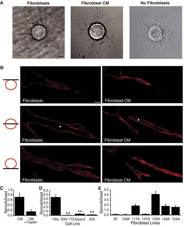
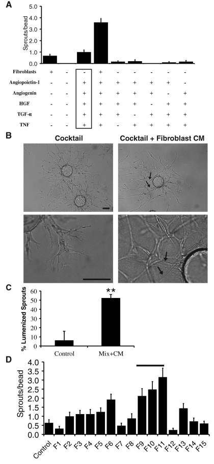
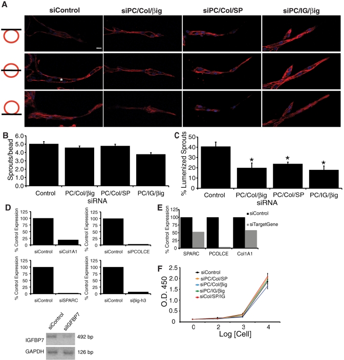
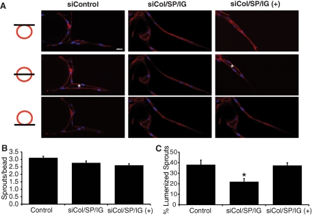
A role for fibroblasts in physiological and pathological angiogenesis is now well recognized; however, the precise mechanisms underlying their action have not been determined. Using an in vitro angiogenesis model in combination with a candidate gene approach, column chromatography, and mass spectrometry, we identify two classes of fibroblast-derived factors--one that supports vessel sprouting but not lumen formation, and one that promotes lumen formation. In the absence of fibroblasts a combination of angiopoietin-1, angiogenin, hepatocyte growth factor, transforming growth factor-α, and tumor necrosis factor drives robust endothelial cell (EC) sprouting; however, lumens fail to form. Subsequent addition of fibroblast-conditioned medium restores lumenogenesis. Using small interfering RNA-mediated knockdown, we show that five genes expressed in fibroblasts--collagen I, procollagen C endopeptidase enhancer 1, secreted protein acidic and rich in cysteine, transforming growth factor-β-induced protein ig-h3, and insulin growth factor-binding protein 7--are necessary for lumen formation. Moreover, lumen formation can be rescued by addition of purified protein to knockdown cultures. Finally, using rheology, we demonstrate that the presence of these matricellular proteins results in significantly stiffer gels, which correlates with enhanced lumen formation. These findings highlight the critical role that fibroblast-derived extracellular matrix components play in EC lumen formation and provide potential insight into the role of fibroblasts in the tumor microenvironment.


Figures

FIGURE 1:
FIGURE 1:
Fibroblasts and fibroblast-CM support EC sprouting and lumen formation, but tumor cells do not. (A) Representative images of fibrin gel bead assays in the presence or absence of fibroblasts and in the presence of fibroblast-CM. Arrows, EC lumens. Scale bar, 50 μm. (B) Confocal images of EC sprouts in the presence of fibroblasts or fibroblast-CM. Three images in three different z-planes for a single sprout are shown. Asterisks indicate EC lumenal space. Scale bar, 10 μm. (C) Quantification of EC sprouting in the fibrin gel bead assay in the presence of fibroblast-CM or fibroblast-CM treated with trypsin. (D) Quantification of ECs sprouting in the fibrin gel bead assay in the presence of fibroblasts or various tumor cell lines. (E) Quantification of EC sprouting in the fibrin gel bead assay in the presence of various fibroblast lines. Data are shown as mean number of sprouts/bead ± standard error of the mean (SEM; n = 20). *p < 0.05; **p < 0.005.
FIGURE 2:
FIGURE 2:
A protein mixture supports EC sprouting but not lumen formation. (A) Quantification of EC sprouting with various combinations of fibroblasts and proteins. Angiogenic cocktail is indicated by the box. Data are shown as mean number of EC sprouts/bead ± SEM (n = 20). (B) Representative images of fibrin gel bead assay. Arrow, EC lumen. Scale bar, 100 μm. (C) Quantification of EC lumen formation in the fibrin gel bead assay. Data are shown as mean percentage of lumenized sprouts/bead ± SEM (n = 20). **p > 0.005. (D) Quantification of EC sprouting in the presence of different fibroblast-CM fractions. Bar, fractions that were pooled for active fraction.
FIGURE 3:
FIGURE 3:
Collagen 1, PCOLCE, SPARC, IGFBP7, and βig-h3 are required for EC lumen formation. (A) Three confocal images in three different z-planes for a single sprout. Asterisks, EC lumenal space. Scale bar, 10 μm. Quantification of EC sprouting (B) and lumen formation (C) in the fibrin gel bead assay in the presence of fibroblasts treated with control siRNA or siRNA targeted to the indicated genes. (D) Relative mRNA levels of targeted gene in fibroblasts treated with the indicated siRNA. (E) Relative mRNA levels of the indicated gene in fibroblasts treated with siRNAs to Col1A1, PCOLCE, and SPARC. (F) XTT cell viability assay. Results in B and C are shown as mean number of EC sprouts/bead or mean percentage of lumenized sprouts/bead, as indicated, ± SEM (n = 60). *p < 0.05.
FIGURE 4:
FIGURE 4:
Addition of exogenous proteins rescues EC lumen formation. (A) Representative confocal images of EC sprouts and lumens in the presence of fibroblasts treated with control siRNA or siRNAs targeted to the indicated genes are shown in the presence or absence of exogenous proteins. Three images in three different z-planes for a single sprout are shown. Asterisks, EC lumenal space. Scale bar, 10 μm. Quantification of EC sprouting (B) and lumen formation (C). Data are shown as mean number of EC sprouts/bead or mean percentage of lumenized sprouts/bead, as indicated, ± SEM (n = 60). *p < 0.05.
FIGURE 5:
FIGURE 5:
Addition of exogenous collagen 1, SPARC, and IGFBP7 increase matrix stiffness. (A) Shear storage modulus of 2.5 mg/ml gels with collagen 1, SPARC, and IGFBP7 or BSA added. Data shown as mean ± SEM (n = 8). ***p < 0.0005. Quantification of EC sprouting (B) and lumen formation (C) in the fibrin gel bead assay in the presence of collagen 1, SPARC, and IGFBP7 or BSA. Data shown as mean ± SEM (n = 30). *p < 0.05.

Similar articles

Cited by

References

    1. Aitkenhead M, Wang SJ, Nakatsu MN, Mestas J, Heard C, Hughes CC. Identification of endothelial cell genes expressed in an in vitro model of angiogenesis: induction of ESM-1, (beta)ig-h3, and NrCAM. Microvasc Res. 2002;63:159–171. - PubMed
    1. Albini A, Magnani E, Noonan DM. The tumor microenvironment: biology of a complex cellular and tissue society. Q J Nucl Med Mol Imaging. 2010;54:244–248. - PubMed
    1. Antoniades HN, Galanopoulos T, Neville-Golden J, Kiritsy CP, Lynch SE. Injury induces in vivo expression of platelet-derived growth factor (PDGF) and PDGF receptor mRNAs in skin epithelial cells and PDGF mRNA in connective tissue fibroblasts. Proc Natl Acad Sci USA. 1991;88:565–569. - PMC - PubMed
    1. Bayless KJ, Salazar R, Davis GE. RGD-dependent vacuolation and lumen formation observed during endothelial cell morphogenesis in three-dimensional fibrin matrices involves the alpha(v)beta(3) and alpha(5)beta(1) integrins. Am J Pathol. 2000;156:1673–1683. - PMC - PubMed
    1. Berthod F, Germain L, Tremblay N, Auger FA. Extracellular matrix deposition by fibroblasts is necessary to promote capillary-like tube formation in vitro. J Cell Physiol. 2006;207:491–498. - PubMed

Publication types

MeSH terms