. 2011 Oct;22(20):3758-67.
doi: 10.1091/mbc.E11-04-0296. Epub 2011 Aug 24.

Mitochondrial Ccs1 contains a structural disulfide bond crucial for the import of this unconventional substrate by the disulfide relay system

Affiliations

Mitochondrial Ccs1 contains a structural disulfide bond crucial for the import of this unconventional substrate by the disulfide relay system

Dominik P Gross et al. Mol Biol Cell. 2011 Oct.

Abstract

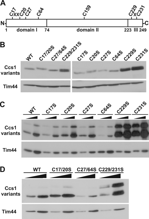
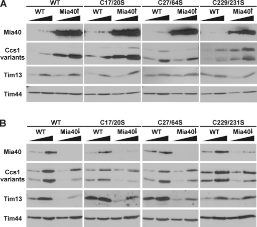
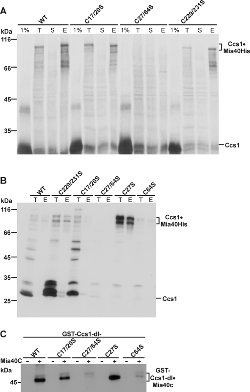
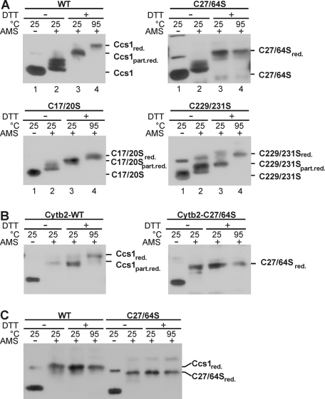
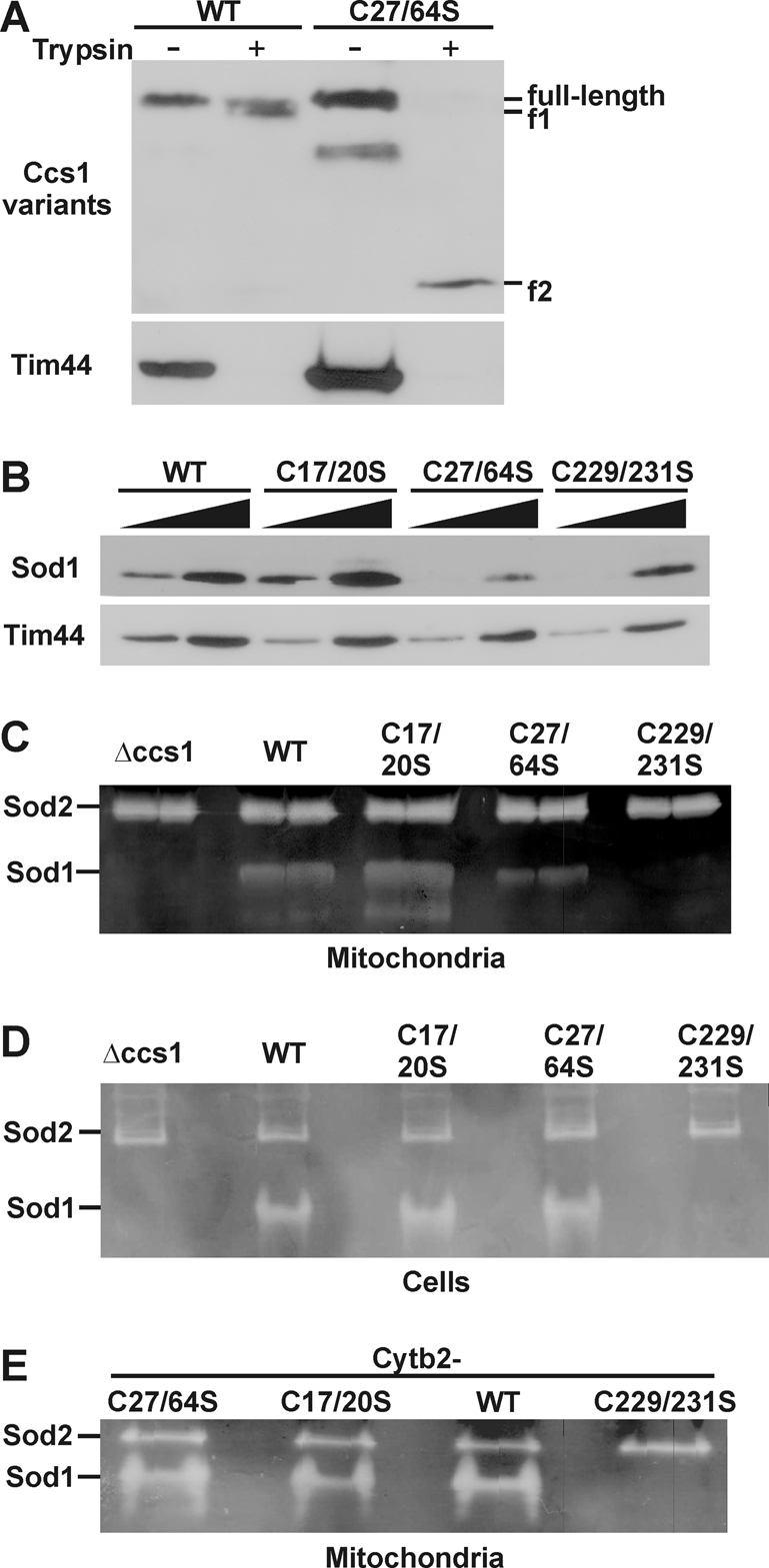
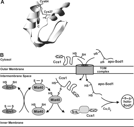
The copper chaperone for superoxide dismutase 1 (Ccs1) provides an important cellular function against oxidative stress. Ccs1 is present in the cytosol and in the intermembrane space (IMS) of mitochondria. Its import into the IMS depends on the Mia40/Erv1 disulfide relay system, although Ccs1 is, in contrast to typical substrates, a multidomain protein and lacks twin Cx(n)C motifs. We report on the molecular mechanism of the mitochondrial import of Saccharomyces cerevisiae Ccs1 as the first member of a novel class of unconventional substrates of the disulfide relay system. We show that the mitochondrial form of Ccs1 contains a stable disulfide bond between cysteine residues C27 and C64. In the absence of these cysteines, the levels of Ccs1 and Sod1 in mitochondria are strongly reduced. Furthermore, C64 of Ccs1 is required for formation of a Ccs1 disulfide intermediate with Mia40. We conclude that the Mia40/Erv1 disulfide relay system introduces a structural disulfide bond in Ccs1 between the cysteine residues C27 and C64, thereby promoting mitochondrial import of this unconventional substrate. Thus the disulfide relay system is able to form, in addition to double disulfide bonds in twin Cx(n)C motifs, single structural disulfide bonds in complex protein domains.


Figures

FIGURE 1:
FIGURE 1:
Distinct cysteine residues in Ccs1 determine the cellular localization of Ccs1. (A) Schematic overview of the domains of S. cerevisiae Ccs1 and of the position of its cysteine residues. III, domain III. (B) Total cell extracts were prepared from cells expressing the indicated cysteine-to-serine exchange variants and wild-type (WT) Ccs1. Cellular proteins were analyzed by SDS–PAGE and immunoblotting with antibodies against Ccs1 and Tim44. Tim44 was used as a control for equal amounts of proteins loaded. (C, D) Mitochondria (12.5, 25 μg) were isolated from cells expressing the indicated single (C) and double (D) cysteine variants of Ccs1 and WT Ccs1. Mitochondrial proteins were analyzed as described earlier. The Ccs1 proteins were expressed with two HA tags. The faster-migrating form of Ccs1 in C and D was not detectable with antibodies against the HA tag (unpublished data), suggesting that these tags are prone to proteolytic removal.
FIGURE 2:
FIGURE 2:
The Mia40-dependent import of Ccs1 depends on distinct cysteine residues. Mitochondria were isolated from cells (A) overexpressing Mia40 (Mia40↑) or (B) depleted of Mia40 (Mia40↓) and from the corresponding wild-type cells (WT). Isolated mitochondria, 12.5 and 25 μg, were analyzed by SDS–PAGE and immunodecoration with antibodies against the indicated proteins. Different times of exposure were chosen for the Ccs1 variants to allow best comparison of the protein levels in WT and Mia40↑, as well as in WT and Mia40↓ mitochondria. Mia40 was more than eightfold overexpressed in Mia40↑ mitochondria. On down-regulation, Mia40 was depleted to at least 10% of the amount present in wild type. The depletion was less prominent in the strain harboring the Ccs1 C229/231S variant. This might explain why the levels of the known Mia40 substrate Tim13, used as a control, were not yet decreased in this mutant, in contrast to the levels in Mia40↓ mitochondria harboring the other Ccs1 variants. However, the levels of Ccs1-C229/231S variant were already reduced. As previously reported, Tim13, present solely in mitochondria, was not affected by the increased Mia40 levels (Reddehase et al., 2009). Tim44, a control for mitochondrial proteins, was present in similar amounts in all mitochondria.
FIGURE 3:
FIGURE 3:
Ccs1 forms a disulfide intermediate via its cysteine residue C64 with Mia40. (A) Radiolabeled precursors of wild-type (WT) Ccs1 and Ccs1 variants were incubated with mitochondria overexpressing Mia40 with an octahistidinyl tag and subsequently treated with IAA. Mitochondria were lysed in Triton X-100–containing buffer and the supernatants were incubated with Ni-NTA agarose beads. Subsequently, beads were washed and bound proteins were eluted. Samples were analyzed by nonreducing SDS–PAGE and autoradiography. It should be noted that the import efficiency was low (<1%). The signal of imported monomeric Ccs1, therefore, could not be distinguished from the unspecific background signal obtained in the import assays. One percent of the amount of radiolabeled precursors used was loaded as input control. E, bound material (100%); S, unbound material (10%); T, total material after lysis (10%). (B) Mitochondria expressing Mia40 with an octahistidinyl tag and the indicated Ccs1 variants were treated with IAA. Mitochondria were lysed, and the extracts were incubated with Ni-NTA agarose beads. Total and bound material was analyzed by SDS–PAGE and immunodecoration with antibodies against Ccs1. (C) The GST-tagged recombinant domains I (Ccs1-dI) of the indicated Ccs1 variants were incubated together with the C-terminal fragment of Mia40 (Mia40C, amino acid residues 284–403) and treated with IAA. Samples were analyzed by nonreducing SDS–PAGE and immunodecoration with antibodies against Mia40.
FIGURE 4:
FIGURE 4:
The cysteine residues C27 and C64 form a disulfide bond. (A) Mitochondria were isolated from cells expressing Ccs1 wild-type (WT) and variants of Ccs1. Mitochondria were incubated for 10 min in absence or presence of 15 mM DTT at 25 or 95°C. Proteins were precipitated with trichloroacetic acid, resuspended in buffer containing 2% SDS, and treated with 10 mM AMS or, when indicated (−AMS), left untreated. Samples were analyzed by SDS–PAGE and immunodecoration with antibodies against Ccs1. Different times of exposure were taken showing the distinct Ccs1 variants. part. red., partially reduced; red., reduced. (B, C) Cells harboring the indicated Ccs1 variants as fusion proteins with the cytochrome b2 targeting signal (B) or the Ccs1 variants (C) were used. Cellular (B) and cytosolic (C) extracts were prepared and samples were treated and analyzed as in A.
FIGURE 5:
FIGURE 5:
The Ccs1 cysteine residues C27 and C64 are crucial for mitochondrial localization of Sod1. (A) Mitochondria isolated from the indicated strains were lysed with Triton X-100 and then incubated on ice in the presence or absence of 200 μg/ml trypsin. After blocking of trypsin activity, samples were analyzed by SDS–PAGE and immunodecoration with antibodies against Ccs1. f1, f2 trypsin-resistant fragment of Ccs1. Three times more protein of the C27/64S mitochondria was loaded. (B, C) Mitochondria were isolated from cells expressing wild-type and the double-cysteine variants of Ccs1. Then they were analyzed by SDS–PAGE and immunoblotting with antibodies against Sod1 and Tim44 (B) and tested for Sod activity in a gel activity assay with nitro blue tetrazolium (C). Immunodecoration with antibodies against Tim44, a mitochondrial protein, indicated the presence of equal amounts of mitochondrial proteins loaded. (D) Activity of Sod1 was analyzed as earlier in extracts from cells expressing the indicated Ccs1 variants. (E) Mitochondria isolated from cells expressing fusion proteins of the cytochrome b2–targeting signal and Ccs1 variants were tested for Sod1 activity as described in C.
FIGURE 6:
FIGURE 6:
Model of the Mia40/Erv1 disulfide relay system–dependent import of Ccs1 and Sod1 into the IMS of mitochondria. (A) Structural representation in ribbon form of the secondary structure of amino acid residues 14–69 of Ccs1. The picture was generated using Swiss-PDB Viewer and PDB file 1qup (Lamb et al., 1999). (B) Ccs1 crosses the TOM complex in the outer membrane of mitochondria in an unfolded state. Following translocation, Ccs1 interacts with oxidized Mia40, forming a disulfide intermediate employing the cysteine residue 64 of its amino-terminal domain. The intermolecular disulfide bond between Mia40 and Ccs1 is then transferred to Ccs1, thereby forming a stable intramolecular disulfide bond between cysteines C27 and C64 that probably promotes folding of Ccs1. On formation of oxidized Ccs1, Mia40 is released in its reduced form and is subsequently reoxidized by Erv1, regenerating oxidized Mia40. Folded Ccs1 mediates mitochondrial import of Sod1, which passes the TOM complex in its reduced apo form. Ccs1 activates apo-Sod1 to holo-Sod1, thereby trapping Sod1 in the IMS. The C-terminal CxC motif of Ccs1, but not the disulfide bond between C27 and C64, is needed for this activation. Copper and oxygen appear to be required as well. The thiol groups of C27 and C64 of Ccs1 are shown in the model.

Similar articles

Cited by

References

    1. Allen S, Balabanidou V, Sideris DP, Lisowsky T, Tokatlidis K. Erv1 mediates the Mia40-dependent protein import pathway and provides a functional link to the respiratory chain by shuttling electrons to cytochrome c. J Mol Biol. 2005;353:937–944. - PubMed
    1. Arnesano F, Balatri E, Banci L, Bertini I, Winge DR. Folding studies of Cox17 reveal an important interplay of cysteine oxidation and copper binding. Structure. 2005;13:713–722. - PubMed
    1. Banci L, Bertini I, Cefaro C, Ciofi-Baffoni S, Gallo A, Martinelli M, Sideris DP, Katrakili N, Tokatlidis K. MIA40 is an oxidoreductase that catalyzes oxidative protein folding in mitochondria. Nat Struct Mol Biol. 2009;16:198–206. - PubMed
    1. Banci L, et al. Molecular chaperone function of Mia40 triggers consecutive induced folding steps of the substrate in mitochondrial protein import. Proc Natl Acad Sci USA. 2010;107:20190–20195. - PMC - PubMed
    1. Bien M, Longen S, Wagener N, Chwalla I, Herrmann JM, Riemer J. Mitochondrial disulfide bond formation is driven by intersubunit electron transfer in Erv1 and proofread by glutathione. Mol Cell. 2010;37:516–528. - PubMed

Publication types

MeSH terms

Substances

LinkOut - more resources