. 2011 Sep;121(9):3398-411.
doi: 10.1172/JCI58186. Epub 2011 Aug 25.

Enteric glia are multipotent in culture but primarily form glia in the adult rodent gut

Affiliations

Enteric glia are multipotent in culture but primarily form glia in the adult rodent gut

Nancy M Joseph et al. J Clin Invest. 2011 Sep.

Abstract

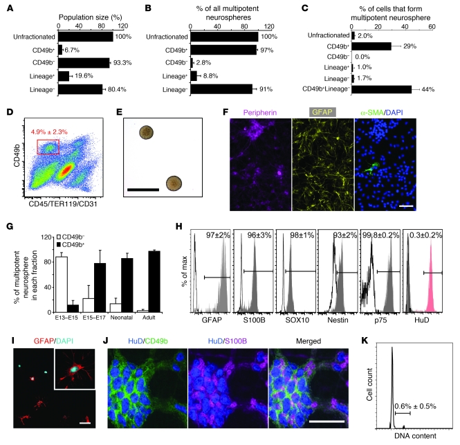
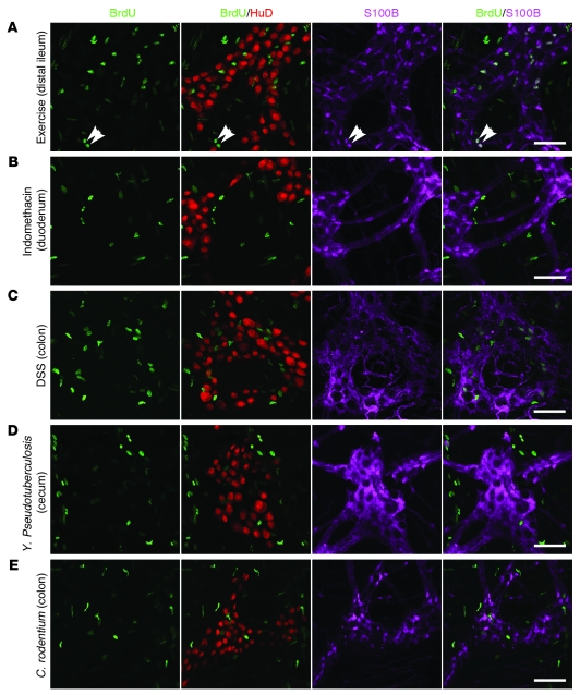
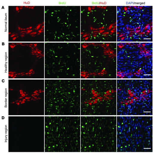
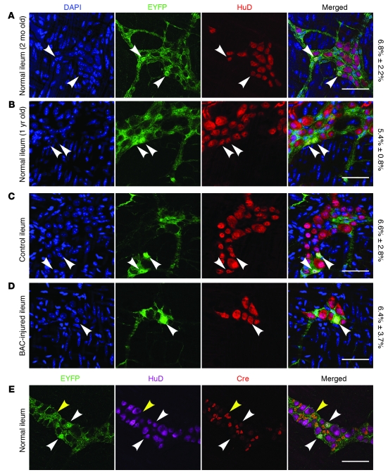
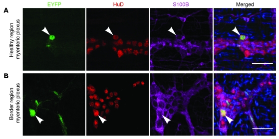
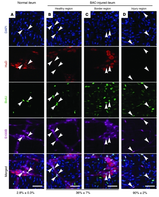
It is unclear whether neurogenesis occurs in the adult mammalian enteric nervous system (ENS). Neural crest-derived cells capable of forming multilineage colonies in culture, and neurons and glia upon transplantation into chick embryos, persist throughout adult life in the mammalian ENS. In this study we sought to determine the physiological function of these cells. We discovered that these cells could be identified based on CD49b expression and that they had characteristics of enteric glia, including p75, GFAP, S100B, and SOX10 expression. To test whether new neurons or glia arise in the adult gut under physiological conditions, we marked dividing progenitors with a thymidine analog in rodents under steady-state conditions, or during aging, pregnancy, dietary changes, hyperglycemia, or exercise. We also tested gut injuries including inflammation, irradiation, benzalkonium chloride treatment, partial gut stenosis, and glial ablation. We readily observed neurogenesis in a neurogenic region of the central nervous system, but not reproducibly in the adult ENS. Lineage tracing of glial cells with GFAP-Cre and GFAP-CreERT2 also detected little or no adult ENS neurogenesis. Neurogenesis in the adult gut is therefore very limited under the conditions we studied. In contrast, ENS gliogenesis was readily observed under steady-state conditions and after injury. Adult enteric glia thus have the potential to form neurons and glia in culture but are fated to form mainly glia under physiological conditions and after the injuries we studied.


Figures

Figure 1
Figure 1. Adult enteric glia form multilineage colonies in culture.
(A) The frequencies of dissociated adult mouse gut cells from the myenteric plexus/muscle layers that stained with antibodies against CD49b or lineage markers (CD45/TER119/CD31). (B) The percentage of all cells that formed multipotent neurospheres that were contained in each sorted cell fraction. (C) The percentage of cells in each sorted fraction that formed multipotent neurospheres in culture. (D) Flow cytometry plot of dissociated adult mouse gut cells stained with antibodies against CD49b and lineage markers. CD49b+Lin cells represented 4.9% ± 2.3% of all unfractionated cells. (E) Primary neurospheres derived from CD49b+Lin cells. (F) These neurospheres generated neurons (peripherin+), glia (GFAP+), and myofibroblasts (α-SMA+). (G) The percentage of all multipotent neurospheres that were formed by CD49b+ or CD49b cells in the fetal, neonatal, and adult mouse guts. (H) Flow-cytometric analysis of fixed CD49b+Lin cells showing that these cells (gray histograms) were nearly uniformly positive for GFAP, S100B, SOX10, nestin, and p75 (unshaded histograms are isotype control staining), but not for the neuronal marker HuD (red shaded histogram represents HuD staining of peripherin+ enteric neurons). (I) GFAP staining of freshly isolated CD49b+Lin cells. (J) CD49b and S100B staining colocalized throughout adult myenteric ganglia, surrounding HuD+ neurons, in a pattern consistent with enteric glia. (K) DNA content of CD49b+Lin cells showing few cells in S/G2/M phases of the cell cycle. Scale bars: 400 μm in E; 50 μm in F, I, and J. All data represent mean ± SD from 3–5 independent experiments.
Figure 2
Figure 2. Voluntary exercise and gut inflammation do not promote neurogenesis in the adult ENS that is detectable by BrdU incorporation.
Whole mount immunostaining of the myenteric plexus with antibodies against BrdU (green), HuD (red), and S100B (magenta) from mice that underwent voluntary exercise (A) or mice with gut inflammation caused by administration of indomethacin (B), DSS (C), Y. pseudotuberculosis (D), or C. rodentium (E). BrdU was administered for 4 months (A) or 6 weeks (BE), and there were 4 mice/treatment. In each treatment we analyzed the distal ileum as well as the stomach and duodenum, colon, or cecum, depending on where the gut inflammation occurred (BE). Gliogenesis was readily observed in all mice (white arrowheads indicating BrdU+S100B+ cells in A; also Supplemental Figure 6). However, we did not observe BrdU+HuD+ neurons in any mice. Most BrdU+ nuclei in all images were from proliferating smooth muscle progenitors and infiltrating inflammatory cells (data not shown). Scale bars: 50 μm.
Figure 3
Figure 3. Focal myenteric plexus ablation by BAC treatment does not promote neurogenesis in the adult ENS that is detectable by BrdU incorporation.
Whole mount staining of myenteric plexus with DAPI (blue) and antibodies against BrdU (green) and HuD (red) in mice after focal ablation of the myenteric plexus by topical BAC. These images were from mice administered BrdU for 4 months, followed by a 1-month chase without BrdU. We analyzed the distal ileum of healthy control mice (A), as well as a healthy region of the gut (B), a region that bordered the ablated region (C), and the ablated region itself (D) in mice treated with BAC. No BrdU+HuD+ cells were observed. We did not observe any neurons within the BAC-ablated region of the myenteric plexus, even 5 months after BAC treatment (D). A total of 70 rats and 15 mice were analyzed in various treatments (see Table 1 for details) that involved BrdU administration for 10 days to 19 weeks. We detected BrdU+HuD+ neurons in only a single rat (see Supplemental Figure 7). Scale bars: 50 μm. The elongated nuclei with more diffuse DAPI staining are from smooth muscle cells in the circular and longitudinal muscle layers surrounding the myenteric plexus.
Figure 4
Figure 4. Lineage tracing in GFAP-Cre;Rosa-loxpEYFP mice failed to detect clear evidence of ENS neurogenesis in healthy or BAC-treated adult mice.
To assess whether new neurons arise from GFAP+ enteric glia during adulthood, we performed lineage tracing in GFAP-Cre;Rosa-loxpEYFP mice. EYFP+HuD+ neurons were counted in healthy young (A; 2 months) and old (B; 12 months) mice as well as in mice treated with topical applications of saline (C) or BAC (D). Three mice were analyzed per treatment, with more than 1,000 HuD+ neurons analyzed per mouse. The numbers on the right represent the percentage of HuD+ neurons that were EYFP+ from each treatment (mean ± SD). BAC-treated mice were analyzed 6–8 months after treatment. (AD) EYFP+HuD+ enteric neurons (white arrowheads) were observed in the myenteric plexus of all mice in all treatments. However, the frequency of EYFP+ neurons did not increase with age (B) or in regions bordering on BAC-ablated areas (D) compared with normal young adult mice (A) or control mice (C), respectively. EYFP+ neurons therefore arose in GFAP-Cre;Rosa-loxpEYFP mice prior to 2 months of age, but few new neurons were generated from GFAP+ cells in the adult ENS. (E) EYFP+ neurons (white arrowheads) did not stain positive for Cre recombinase, while EYFP+ glia did (yellow arrowheads), suggesting that EYFP expression was not due to nonspecific expression of Cre recombinase in enteric neurons. Scale bars: 50 μm.
Figure 5
Figure 5. Lineage tracing in adult GFAP-CreERT2;Rosa-loxpEYFP mice detected little ENS neurogenesis after BAC treatment.
To assess whether new neurons arise from GFAP+ enteric glia during adulthood, we treated GFAP-CreERT2;Rosa-loxpEYFP mice with BAC to ablate myenteric neurons and then with tamoxifen for 4–8 months. EYFP+HuD+ neurons (white arrowheads) were counted in a healthy region of the gut (A), as well as in a border region immediately adjacent to the BAC-ablated area (B). The frequency of EYFP+HuD+ neurons was not increased in the border region immediately adjacent to the BAC-ablated area (B; 0.028% ± 0.027%, see Table 2) as compared with a healthy segment of gut in the same mice (A; 0.030% ± 0.022%, see Table 2). It is not clear whether this reflects the generation of rare neurons in the adult ENS in a manner that is not influenced by BAC injury, or the ectopic expression of Cre recombinase in rare neurons. Scale bars: 50 μm.
Figure 6
Figure 6. Gliogenesis occurs in the adult ENS under steady-state conditions and in response to injury.
Whole mount staining of the myenteric plexus with DAPI (blue) and with antibodies against HuD (red), BrdU (green), and S100B (magenta) from normal adult mice (A) and mice that underwent focal ablation of the myenteric plexus by topical BAC treatment (BD). BrdU was administered for 6 weeks, followed by a chase without BrdU for 6 weeks in A; and administered for 10 weeks, followed by a chase without BrdU for 3 weeks in BD. Three mice were analyzed per treatment, with 2,000–4,000 glial cells counted per mouse. (A) 2.8% ± 0.3% of S100B+ glial cells in the myenteric plexus of healthy adult mice were BrdU+ after 6 weeks of BrdU treatment, indicating that gliogenesis does occur under steady-state conditions in the adult ENS. (B and C) Compared with uninjured mice, we observed increased frequencies (36% ± 7%) of S100B+BrdU+ glia in the myenteric plexus of BAC-treated mice, in the region bordering the injury and in healthy regions several inches upstream of the injury. (D) Almost all (90% ± 2%) S100B+ glia within the BAC-ablated region were BrdU+, suggesting a regenerative response to restore glia to the ablated region. Numbers on the bottom indicate the frequency of S100B+ myenteric plexus glia that were BrdU+ (mean ± SD). Scale bars: 50 μm.

Comment in

References

    1. Altman J, Das GD. Autoradiographic and histological evidence of postnatal hippocampal neurogenesis in rats. J Comp Neurol. 1965;124(3):319–335. doi: 10.1002/cne.901240303. - DOI - PubMed
    1. Altman J, Das GD. Autoradiographic and histological studies of postnatal neurogenesis. J Comp Neurol. 1966;126(3):337–390. doi: 10.1002/cne.901260302. - DOI - PubMed
    1. Altman J. Autoradiographic and histological studies of postnatal neurogenesis. IV. Cell proliferation and migration in the anterior forebrain, with special reference to persisting neurogenesis in the olfactory bulb. J Comp Neurol. 1969;137(4):433–458. doi: 10.1002/cne.901370404. - DOI - PubMed
    1. Luskin MB. Restricted proliferation and migration of postnatally generated neurons derived from the forebrain subventricular zone. Neuron. 1993;11(1):173–189. - PubMed
    1. Seki T, Arai Y. Highly polysialylated neural cell adhesion molecule (NCAM-H) is expressed by newly generated granule cells in the dentate gyrus of the adult rat. J Neurosci. 1993;13(6):2351–2358. - PMC - PubMed

Publication types

MeSH terms