. 2011 Sep;121(9):3589-97.
doi: 10.1172/JCI58447. Epub 2011 Aug 25.

A genetically engineered human pancreatic β cell line exhibiting glucose-inducible insulin secretion

Affiliations

A genetically engineered human pancreatic β cell line exhibiting glucose-inducible insulin secretion

Philippe Ravassard et al. J Clin Invest. 2011 Sep.

Abstract

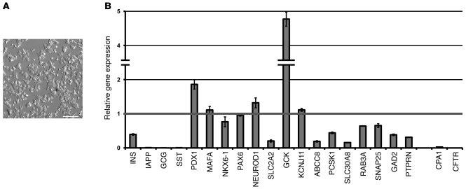
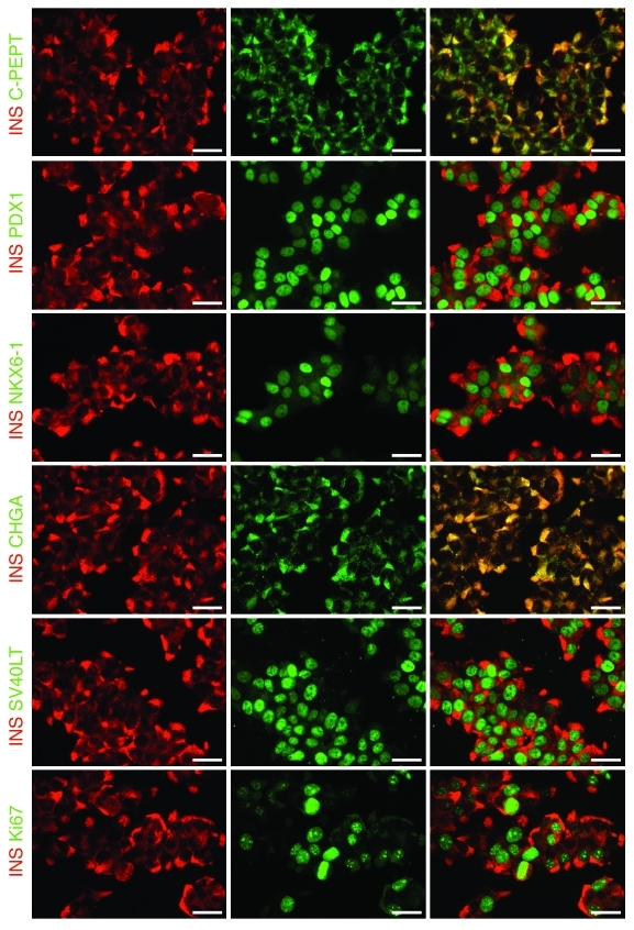
Despite intense efforts over the past 30 years, human pancreatic β cell lines have not been available. Here, we describe a robust technology for producing a functional human β cell line using targeted oncogenesis in human fetal tissue. Human fetal pancreatic buds were transduced with a lentiviral vector that expressed SV40LT under the control of the insulin promoter. The transduced buds were then grafted into SCID mice so that they could develop into mature pancreatic tissue. Upon differentiation, the newly formed SV40LT-expressing β cells proliferated and formed insulinomas. The resulting β cells were then transduced with human telomerase reverse transcriptase (hTERT), grafted into other SCID mice, and finally expanded in vitro to generate cell lines. One of these cell lines, EndoC-βH1, expressed many β cell-specific markers without any substantial expression of markers of other pancreatic cell types. The cells secreted insulin when stimulated by glucose or other insulin secretagogues, and cell transplantation reversed chemically induced diabetes in mice. These cells represent a unique tool for large-scale drug discovery and provide a preclinical model for cell replacement therapy in diabetes. This technology could be generalized to generate other human cell lines when the cell type-specific promoter is available.


Figures

Figure 1
Figure 1. In vivo expansion of SV40LT-expressing cells in transplanted human fetal pancreas.
Human fetal pancreases that were transduced or not transduced with an SV40LT-expressing lentiviral vector were transplanted under the kidney capsule of SCID mice. 3 months later, insulin (INS) immunostaining (red) revealed β cell clusters in both the nontransduced and SV40LT-transduced fetal pancreases. In the nontransduced pancreases, the β cells stained negative for both SV40LT (green) and Ki67 (green), whereas β cell clusters coexpressed SV40LT and INS in the SV40LT-transduced transplants. Such clusters proliferated, as visualized by Ki67 staining. Scale bars: 50 μm.
Figure 2
Figure 2. Primary insulinomas develop from SV40LT-transduced fetal pancreases, but not from adult islets.
Human fetal pancreases and adult islets were transduced with an SV40LT-expressing lentiviral vector, transplanted under kidney (K) capsule, and then removed 6 to 8 months later. (AD) In the transduced human fetal pancreatic grafts, substantial tissue expansion and highly vascularized regions were observed (arrows). Histological examination of sections of this vascularized domain revealed a large population of cells that stained positively for both SV40LT (green) and insulin (red) and expressed Ki67 (green). (EH) In transduced human adult islet grafts, insulin-positive cells (red) were abundant, and SV40LT-positive cells (green) were rarely observed. However, their proliferation remained low, as measured by Ki67 (green), and no tissue expansion was observed. (A and E) Square size in grids: 1 cm. Scale bars: 50 μm.
Figure 3
Figure 3. Secondary insulinomas are homogeneously composed of proliferating insulin-positive cells.
Primary insulinomas were transduced with a lentiviral vector that expressed hTERT and then transplanted into SCID mice. 2 months later, secondary insulinomas were analyzed by immunostaining. (AE) The entire tissue stained positive for insulin (INS) (red). All nuclei (blue) were positive for SV40LT (A, green) and PDX1 (C, green). A large number of cells proliferated, as indicated by Ki67-positive staining (B, green). No expression of glucagon (D) or amylase (E) was detected. Scale bars: 50 μm.
Figure 4
Figure 4. Morphology and gene expression profiling of EndoC-βH1 cells.
(A) Morphology of EndoC-βH1 cells at passage 63. Scale bar: 250 μm (B) Quantitative real-time PCR analyses of the indicated genes were performed using cDNA prepared from 3 independent cultures of EndoC-βH1 cells and from 1 adult human islet preparation. Results are presented as mean ± SEM of the levels that were measured in human islets.
Figure 5
Figure 5. Immunofluorescence analysis of EndoC-βH1 cells.
EndoC-βH1 cells stained positive for insulin and coexpressed C-peptide, PDX1, NKX6-1, CHGA, SV40LT, and Ki67. Scale bars: 25 μm.
Figure 6
Figure 6. EndoC-βH1 cells secrete insulin in vitro.
(A) EndoC-βH1 cells secreted insulin in response to glucose stimulation. (B) Insulin secretion by EndoC-βH1 cells increased when stimulated with 100 nM exendin-4 (EX4), 100 μM glibenclamide (Gli), 500 μM IBMX, and 10 mM l-leucine (Leu). Diazoxine (DZ) treatment (500 μM) reduced insulin secretion to basal levels. (C) Insulin secretion was first stimulated for 60 minutes with 11 mM glucose in the presence or absence of IBMX. This medium was the replaced with HEPES-buffered Krebs-Ringer Buffer that contained 2.8 mM glucose, and the cells were further incubated in this medium for 60 minutes. Note that insulin secretion returned to basal level in the presence of 2.8 mM glucose. The figure shows data from a representative experiment that was done in duplicate on 3 experimental replicates. Results are expressed as the mean percentage ± SEM of the insulin content that was secreted in 1 hour.
Figure 7
Figure 7. Normoglycemia is restored after an EndoC-βH1 cell transplant into diabetic SCID mice.
(A) DM was chemically induced in 12 SCID mice by an intraperitoneal injection of STZ. 2 days later, 2 × 10⁶ EndoC-βH1 cells were transplanted under the kidney capsule in 6 of the 12 DM mice. The transplanted cells were removed by unilateral nephrectomy 55 days later. Each point is the mean ± SEM of blood glucose levels that were measured at different time points in the transplanted (circles) or nontransplanted (squares) SCID mice. (B) An intraperitoneal glucose tolerance test was performed on the STZ-induced DM SCID mice with the EndoC-βH1 cell graft, 35 days after the cell transplant. Proper regulation of blood glucose levels was observed in the cell-transplanted DM SCID mice (circles) when compared with those of the control non-DM SCID mice (squares). Each point in the graph is the mean ± SEM from 4 mice in each group. (C) Insulin (red) and Ki67 (green) costaining in the cell transplant after its removal by unilateral nephrectomy. (D) The pancreas of the DM mice (+STZ) with the EndoC-βH1 cell graft is almost devoid of insulin-positive cells (brown), whereas non-DM mice (–STZ) present a large number of insulin-positive islets. Scale bars: 25 μm (C); 500 μm (100 μm in the inset) (D).

Comment in

  • Finally! A human pancreatic β cell line.
    Weir GC, Bonner-Weir S. Weir GC, et al. J Clin Invest. 2011 Sep;121(9):3395-7. doi: 10.1172/JCI58899. Epub 2011 Aug 25. J Clin Invest. 2011. PMID: 21865653 Free PMC article.

Similar articles

Cited by

References

    1. Beattie GM, et al. Sustained proliferation of PDX-1+ cells derived from human islets. Diabetes. 1999;48(5):1013–1019. doi: 10.2337/diabetes.48.5.1013. - DOI - PubMed
    1. Russ HA, Bar Y, Ravassard P, Efrat S. In vitro proliferation of cells derived from adult human beta-cells revealed by cell-lineage tracing. Diabetes. 2008;57(6):1575–1583. doi: 10.2337/db07-1283. - DOI - PubMed
    1. Kroon E, et al. Pancreatic endoderm derived from human embryonic stem cells generates glucose-responsive insulin-secreting cells in vivo. Nat Biotechnol. 2008;26(4):443–452. doi: 10.1038/nbt1393. - DOI - PubMed
    1. Asfari M, Janjic D, Meda P, Li G, Halban P, Wolheim K. Establishement of 2-mercaptoethanol-dependent differentiated insulin secreting cell lines. Endocrinology. 1992;130(1):167–178. doi: 10.1210/en.130.1.167. - DOI - PubMed
    1. Gazdar A, et al. Continuous, clonal, insulin- and somatostatin-secreting cell lines established from a transplantable rat islet cell tumor. Proc Natl Acad Sci U S A. 1980;77(6):3519–3523. doi: 10.1073/pnas.77.6.3519. - DOI - PMC - PubMed

Publication types