. 2011 Sep;121(9):3701-12.
doi: 10.1172/JCI44736. Epub 2011 Aug 25.

Parkin is a lipid-responsive regulator of fat uptake in mice and mutant human cells

Affiliations

Parkin is a lipid-responsive regulator of fat uptake in mice and mutant human cells

Kye-Young Kim et al. J Clin Invest. 2011 Sep.

Abstract

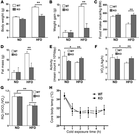
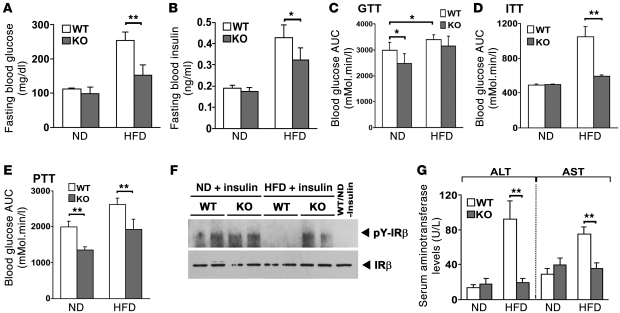
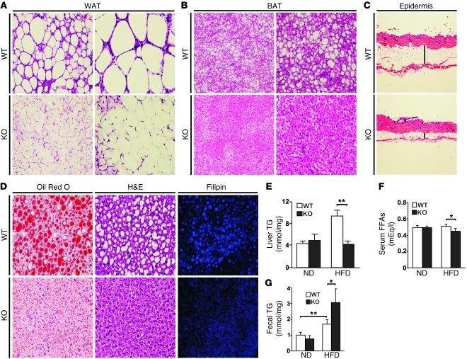
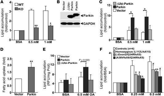
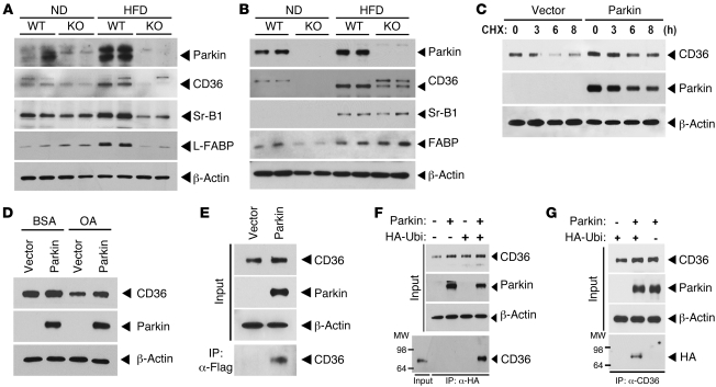
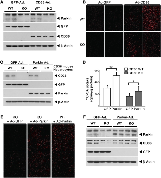
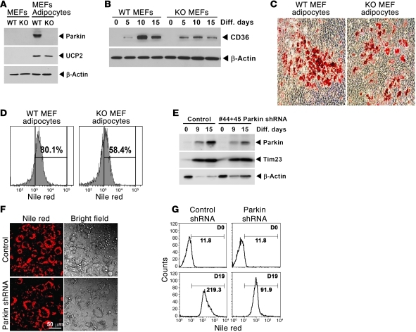
It has long been hypothesized that abnormalities in lipid biology contribute to degenerative brain diseases. Consistent with this, emerging epidemiologic evidence links lipid alterations with Parkinson disease (PD), and disruption of lipid metabolism has been found to predispose to α-synuclein toxicity. We therefore investigated whether Parkin, an E3 ubiquitin ligase found to be defective in patients with early onset PD, regulates systemic lipid metabolism. We perturbed lipid levels by exposing Parkin+/+ and Parkin-/- mice to a high-fat and -cholesterol diet (HFD). Parkin-/- mice resisted weight gain, steatohepatitis, and insulin resistance. In wild-type mice, the HFD markedly increased hepatic Parkin levels in parallel with lipid transport proteins, including CD36, Sr-B1, and FABP. These lipid transport proteins were not induced in Parkin-/- mice. The role of Parkin in fat uptake was confirmed by increased oleate accumulation in hepatocytes overexpressing Parkin and decreased uptake in Parkin-/- mouse embryonic fibroblasts and patient cells harboring complex heterozygous mutations in the Parkin-encoding gene PARK2. Parkin conferred this effect, in part, via ubiquitin-mediated stabilization of the lipid transporter CD36. Reconstitution of Parkin restored hepatic fat uptake and CD36 levels in Parkin-/- mice, and Parkin augmented fat accumulation during adipocyte differentiation. These results demonstrate that Parkin is regulated in a lipid-dependent manner and modulates systemic fat uptake via ubiquitin ligase-dependent effects. Whether this metabolic regulation contributes to premature Parkinsonism warrants investigation.


Figures

Figure 1
Figure 1. Parkin–/– mice (KO) are resistant to HFD-induced weight gain and have preserved activity and oxygen consumption.
12-week-old male mice were fed a ND or a HFD. BW (A) and weight gain (B) measured after 6.5 weeks of the ND compared with the HFD. (C) Daily food intake (kcal/kg BW) was measured for 3 days while mice were housed in a metabolic chamber. The mice were fed a ND or a HFD for 5 weeks prior to analysis. (D) Total body fat mass was analyzed by NMR spectroscopy. (E) Daily total movement counted by DSI telemetry system and averaged over 3 days. (F) Total metabolic rate (O2 consumption) monitored by Oxymax system for 24 hours. VO2, volume of O2 consumed. (G) Respiratory quotient (RQ) measured for 24 hours. VCO2, volume of CO2 consumed. (H) Core body temperature response to a hypothermia challenge (4°C for 5 hours). Data are expressed as mean ± SD (n = 5–9 per group). *P < 0.05; **P < 0.01, compared with the corresponding controls.
Figure 2
Figure 2. Parkin–/– mice are resistant to HFD-induced glucose intolerance and hepatic insulin resistance.
(A and B) Fasting blood glucose and serum insulin levels in Parkin+/+ and Parkin–/– mice. (C) GTT expressed as the AUC of blood glucose levels during the 2 hour study. (D) ITT expressed as AUC for blood glucose over 2 hours. (E) PTT expressed as AUC for blood glucose of 2 hours. (F) Assessment of phosphorylation of IRβ in liver following intraperitoneal insulin administration. Liver extracts were IP with IRβ antibody and the immunoblot performed with a phospho-tyrosine antibody. The far right lane shows protein expression in WT mice on the ND without insulin. (G) Serum aminotransferase levels. The mice were fed with ND or HFD for 6.5 weeks. Data are expressed as mean ± SD. *P < 0.05; **P < 0.01 versus controls (n = 4–6 per group).
Figure 3
Figure 3. Parkin–/– mice are resistant to HFD-induced fat accumulation.
Tissue sections were analyzed after 6.5 weeks of the HFD. WAT (A) and BAT (B) were processed using H&E staining. The upper panels show Parkin+/+ (WT) mice and lower panels show Parkin–/– (KO) mice. Original magnification, ×10 (left panels); ×20 (right panels). (C) The skin epidermal thickness in transverse sections from Parkin+/+ and Parkin–/– mice (H&E staining). Vertical line shows the subcutaneous fat. (D) TG accumulation in liver sections by Oil Red O and H&E staining, and cholesterol levels by Filipin staining in Parkin WT and Parkin–/– mice. Original magnification, ×20 (left panels); ×20 (middle panels); ×40 (right panels).(E) Hepatic TG levels, (F) serum-free fatty acid levels, and (G) TG levels in the feces on the ND and HFD comparing the WT and KO mice. The feces from the mice fed a ND (n = 4) or HFD (n = 7) for 5 weeks were collected daily over a 3-day period. Lipids were extracted from the feces and TG measured using a colorimetric assay. Data are expressed as mean ± SD. Representative histological images are shown; similar results were acquired from 3–4 independent mice. *P < 0.05; **P < 0.01 versus controls (n = 4–7 per group).
Figure 4
Figure 4. Parkin modulation of lipid uptake requires an intact ubiquitin-like domain.
(A) Lipid accumulation assayed by Nile red staining in MEFs from Parkin+/+ (WT) and Parkin–/– (KO) after BSA-conjugated oleate (0.5 and 1 mM) incubation. Values represent the fold relative to BSA incubation and normalized to cell number. The values represent the average of 5 independent experiments. (B) Parkin expression in HepG2 cells overexpressing vector, WT Parkin, and ∆Ubi-Parkin constructs. (C) Lipid accumulation in HepG2 cells overexpressing vector, Parkin, and ∆Ubi-Parkin after oleate incubation (0.5 and 1 mM). (D) Relative fluorescence of Bodipy-labeled dodecanoic acid at the end of 1,200-second incubation in HepG2 cells overexpressing Parkin compared with vector controls. Values represent the fold relative to vector-transfected HepG2 cells. (E) Lipid accumulation in SH-SY5Y neuroblastoma cells overexpressing vector, Parkin, and ΔUbi-Parkin after 0.5 mM oleate incubation. Values are normalized to protein concentration. (F) Lipid accumulation in response to 0.25 and 0.5 mM oleate in transformed B cells from 3 patients with PARK2 mutations versus 4 WT control subjects. Values were normalized to cell number. Data are displayed relative to control cells exposed to BSA normalized to 1. Data are expressed as mean ± SD. *P < 0.05; **P < 0.01 versus control or vector.
Figure 5
Figure 5. Parkin regulates posttranslational modification of CD36.
(A) Expression levels of Parkin and lipid uptake (CD36 and Sr-B1) and transport (L-FABP) proteins in response to the ND and HFD in liver tissue by immunoblot analysis. (B) Protein expression of Parkin and lipid uptake and transport proteins in response to the ND and HFD in whole brain tissue by immunoblot analysis. (C) Degradation of CD36 protein following CHX administration in the presence or absence of Parkin overexpression in HeLa cells engineered to overexpress CD36. (D) CD36 protein expression in response to oleic acid administration following overexpression of Parkin or control vector. (E) Interaction between Parkin and CD36. CD36-overexpressing HeLa cells were transfected with p3×Flag vector or p3×Flag-Parkin, respectively. After 48 hours transfection, protein extracts were IP with anti-Flag M2 agarose (IP:α-Flag). (F) Ubiquitination of CD36 following IP of an HA antibody and immunoblot analysis for CD36. CD36-overexpressing HeLa cells were transfected with HA-Ubiquitin and/or Parkin. (G) Ubiquitination of CD36 following IP with a CD36 antibody and immunoblot analysis for HA following the transfection of HA-Ubiquitin and/or Parkin in the CD36-overexpressing HeLa cells. All studies were performed in duplicate, and at least 3 independent experiments were performed.
Figure 6
Figure 6. Fat uptake is enhanced following reconstitution of CD36 or Parkin.
(A) Immunoblot analysis showing overexpression of CD36 versus GFP control adenovirus (Ad) in Parkin+/+ and Parkin–/– mice liver. A short exposure of the CD36 immunoblot bands is shown to maintain the viral overexpression levels within the linear range. (B) Confocal microscopy of fixed liver tissue showing enhanced VLDL uptake following CD36 infection (red fluorescence). Representative fat levels are shown in 2 Parkin+/+ and Parkin–/– mice following either GFP or CD36 adenoviral infection. Original magnification, ×40. (C) Immunoblot analysis showing GFP or Parkin expression in Cd36+/+ and Cd36–/– primary hepatocytes following either GFP or Parkin adenoviral infection. (D) Radiolabeled oleate uptake in Cd36+/+ and Cd36–/– primary hepatocytes. Data are expressed as mean ± SD. *P < 0.05; **P < 0.01 versus controls. (E) Confocal microscopy showing enhanced VLDL uptake following Parkin infection in mouse livers. Original magnification, ×40. (F) Immunoblot analysis showing induction of Parkin and CD36 in Parkin WT and Parkin–/– mice on the HFD following infection with Parkin or GFP adenoviral particles.
Figure 7
Figure 7. Parkin is regulated in parallel with fat accumulation in adipocytes and functions to facilitate fat uptake during adipogenesis.
(A) Representative immunoblot showing increased Parkin expression in Parkin+/+ MEF cells on day 21 adipocyte differentiation. The ubiquitously expressed mitochondrial uncoupling protein 2 (UCP2) is increased during adipogenesis in both Parkin+/+ and Parkin–/– MEF cells. (B) Representative immunoblot showing the temporal induction of CD36 levels in Parkin+/+ and Parkin–/– MEFs. β-actin levels reflect protein loading. (C) Light microscopy shows Oil Red O staining on day 21 adipocyte differentiation in MEFs with increased staining in Parkin WT MEFs. Original magnification, ×10. (D) Representative flow cytometric profile showing Nile red uptake in differentiated MEF cells to compare cellular fat accumulation. The percentages shown represent the increase in neutral lipid accumulation above predifferentiated MEF cell levels. (E) Immunoblot analysis of 3T3-L1 cell differentiation with higher Parkin levels in the scrambled shRNA-treated cells versus those transfected with 2 Parkin shRNAs. The inner mitochondrial membrane transport protein (Tim23) is increased during adipogenesis and shows similar expression in control and Parkin shRNA–treated cells. (F) Light microscopy to show fat accumulation in differentiated 3T3-L1 adipocytes by Nile red or by bright-field microscopy comparing control and Parkin shRNA–infected cells. Original magnification, ×20. (G) Flow cytometry showing significantly more neutral lipid accumulation in control versus Parkin shRNA–infected 3T3-L1 cell adipocyte differentiation on day 19 (D19) versus the cytometric profile prior to differentiation (D0). The numbers adjacent to the distribution curves represent the geometric mean of fluorescence intensity in the cell populations gated to measure Nile red accumulation. All experiments were repeated 3 or more times.

Comment in

References

    1. Kitada T, et al. Mutations in the parkin gene cause autosomal recessive juvenile parkinsonism. Nature. 1998;392(6676):605–608. doi: 10.1038/33416. - DOI - PubMed
    1. Goldberg MS, et al. Parkin-deficient mice exhibit nigrostriatal deficits but not loss of dopaminergic neurons. J Biol Chem. 2003;278(44):43628–43635. doi: 10.1074/jbc.M308947200. - DOI - PubMed
    1. Perez FA, Palmiter RD. Parkin-deficient mice are not a robust model of parkinsonism. Proc Natl Acad Sci U S A. 2005;102(6):2174–2179. doi: 10.1073/pnas.0409598102. - DOI - PMC - PubMed
    1. Kitada T, Tong Y, Gautier CA, Shen J. Absence of nigral degeneration in aged parkin/DJ-1/PINK1 triple knockout mice. J Neurochem. 2009;111(3):696–702. - PMC - PubMed
    1. Beasley SA, Hristova VA, Shaw GS. Structure of the Parkin in-between-ring domain provides insights for E3-ligase dysfunction in autosomal recessive Parkinson’s disease. Proc Natl Acad Sci U S A. 2007;104(9):3095–3100. doi: 10.1073/pnas.0610548104. - DOI - PMC - PubMed

Publication types

MeSH terms