

Accumulation of transactive response DNA binding protein 43 in mild cognitive impairment and Alzheimer disease
- PMID: 21865887
- PMCID: PMC3197017
- DOI: 10.1097/NEN.0b013e31822c62cf
Accumulation of transactive response DNA binding protein 43 in mild cognitive impairment and Alzheimer disease
Abstract
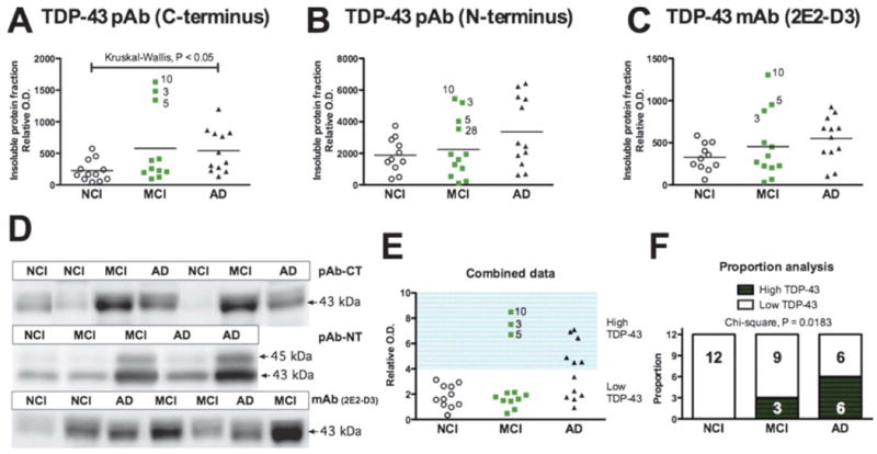
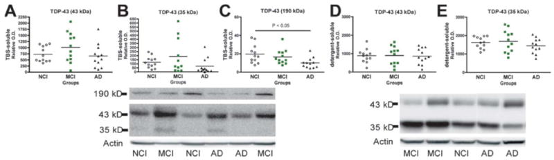
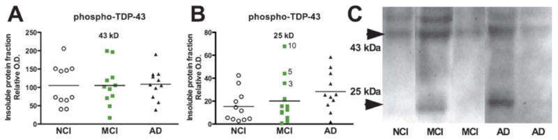
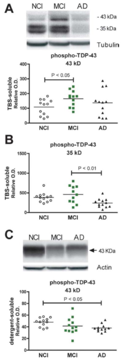
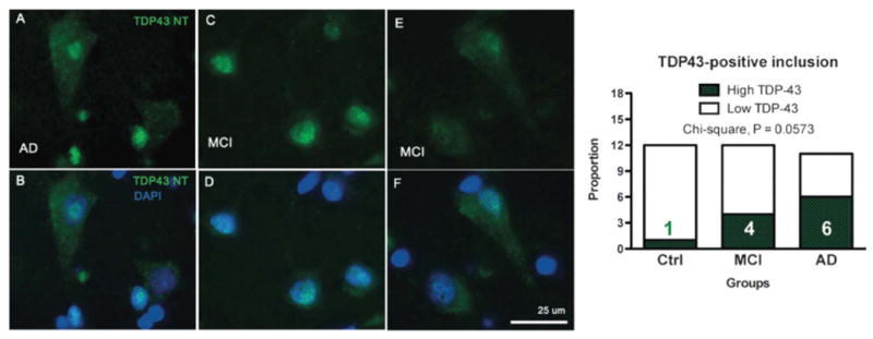
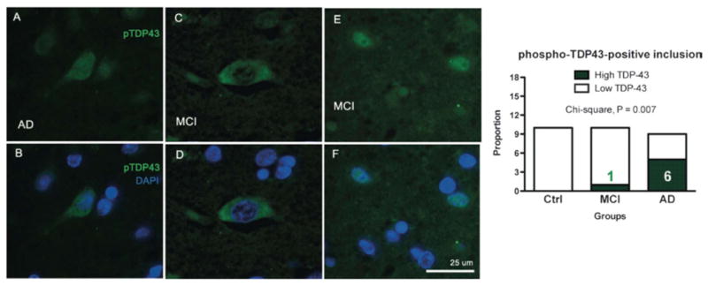
Transactive response DNA binding protein 43 (TDP-43) plays a central role in the neuropathology of frontotemporal lobar degeneration and amyotrophic lateral sclerosis, but the relationship between TDP-43 abnormalities and Alzheimer disease (AD) remains unclear. To determine whether TDP-43 can serve as a neuropathologic marker of AD, we performed biochemical characterization and quantification of TDP-43 in homogenates from parietal neocortex of subjects with aclinical diagnosis of no cognitive impairment (NCI, n = 12), mild cognitive impairment (MCI, n = 12), or AD (n = 12). Immunoblots revealed increased detergent-insoluble TDP-43 in the cortex of 0, 3, and 6 of the 12 individuals with NCI, MCI, or AD, respectively. Detergent-insoluble TDP-43 was positively correlated with the accumulation of soluble Aβ42, amyloid plaques, and paired helical filamenttau. In contrast, phospho-TDP-43 was decreased in the cytosolic fraction and detergent-soluble membrane/nuclear fraction from AD patients and correlated with antemortem cognitive function.Immunofluorescence analysis confirmed that the frequencies of individuals with TDP-43 or phospho-TDP-43 cytoplasmic inclusions were higher in AD than in NCI, with MCI at an intermediate level. These data indicate that abnormalities of TDP-43 occur in an important subset of MCI and AD patients and that they correlate with the clinical and neuropathologic features of AD.
Figures
Similar articles
-
Interaction of transactive response DNA binding protein 43 with nuclear factor κB in mild cognitive impairment with episodic memory deficits.Acta Neuropathol Commun. 2014 Apr 1;2:37. doi: 10.1186/2051-5960-2-37. Acta Neuropathol Commun. 2014. PMID: 24690380 Free PMC article.
-
Biochemical characterization of Abeta and tau pathologies in mild cognitive impairment and Alzheimer's disease.J Alzheimers Dis. 2007 Dec;12(4):377-90. doi: 10.3233/jad-2007-12411. J Alzheimers Dis. 2007. PMID: 18198424
-
Precuneus amyloid burden is associated with reduced cholinergic activity in Alzheimer disease.Neurology. 2011 Jul 5;77(1):39-47. doi: 10.1212/WNL.0b013e3182231419. Epub 2011 Jun 22. Neurology. 2011. PMID: 21700583 Free PMC article.
-
The Role of TDP-43 in Alzheimer's Disease.Mol Neurobiol. 2016 Jul;53(5):3349-3359. doi: 10.1007/s12035-015-9264-5. Epub 2015 Jun 17. Mol Neurobiol. 2016. PMID: 26081142 Review.
-
TDP-43 in aging and Alzheimer's disease - a review.Int J Clin Exp Pathol. 2011 Jan 30;4(2):147-55. Int J Clin Exp Pathol. 2011. PMID: 21326809 Free PMC article. Review.
Cited by
-
The overexpression of TDP-43 in astrocytes causes neurodegeneration via a PTP1B-mediated inflammatory response.J Neuroinflammation. 2020 Oct 14;17(1):299. doi: 10.1186/s12974-020-01963-6. J Neuroinflammation. 2020. PMID: 33054766 Free PMC article.
-
Overlapping but distinct TDP-43 and tau pathologic patterns in aged hippocampi.Brain Pathol. 2018 Mar;28(2):264-273. doi: 10.1111/bpa.12505. Epub 2017 Mar 24. Brain Pathol. 2018. PMID: 28281308 Free PMC article.
-
Postmortem Fatty Acid Abnormalities in the Cerebellum of Patients with Essential Tremor.Cerebellum. 2024 Dec;23(6):2341-2359. doi: 10.1007/s12311-024-01736-4. Epub 2024 Aug 31. Cerebellum. 2024. PMID: 39215908 Free PMC article.
-
Higher Angiotensin I Converting Enzyme 2 (ACE2) levels in the brain of individuals with Alzheimer's disease.bioRxiv [Preprint]. 2023 Jan 18:2023.01.17.524254. doi: 10.1101/2023.01.17.524254. bioRxiv. 2023. Update in: Acta Neuropathol Commun. 2023 Oct 2;11(1):159. doi: 10.1186/s40478-023-01647-1. PMID: 36711734 Free PMC article. Updated. Preprint.
-
Beta-amyloid pathology in human brain microvessel extracts from the parietal cortex: relation with cerebral amyloid angiopathy and Alzheimer's disease.Acta Neuropathol. 2019 May;137(5):801-823. doi: 10.1007/s00401-019-01967-4. Epub 2019 Feb 7. Acta Neuropathol. 2019. PMID: 30729296 Free PMC article.
References
-
- Neumann M, Sampathu DM, Kwong LK, et al. Ubiquitinated TDP-43 in frontotemporal lobar degeneration and amyotrophic lateral sclerosis. Science. 2006;314:130–3. - PubMed
-
- Mackenzie IRA, Bigio EH, Ince PG, et al. Pathological TDP-43 distinguishes sporadic amyotrophic lateral sclerosis from amyotrophic lateral sclerosis with SOD1 mutations. Ann Neurol. 2007;61:427–34. - PubMed
-
- Arai T, Hasegawa M, Akiyama H, et al. TDP-43 is a component of ubiquitin-positive tau-negative inclusions in frontotemporal lobar degeneration and amyotrophic lateral sclerosis. Biochem Biophys Res Commun. 2006;351:602–11. - PubMed
Publication types
MeSH terms
Substances
Grants and funding
LinkOut - more resources
Full Text Sources
Other Literature Sources
Medical
