. 2011 Aug 24;477(7362):61-6.
doi: 10.1038/nature10362.

The mechanism of membrane-associated steps in tail-anchored protein insertion

Affiliations

The mechanism of membrane-associated steps in tail-anchored protein insertion

Malaiyalam Mariappan et al. Nature. .

Abstract

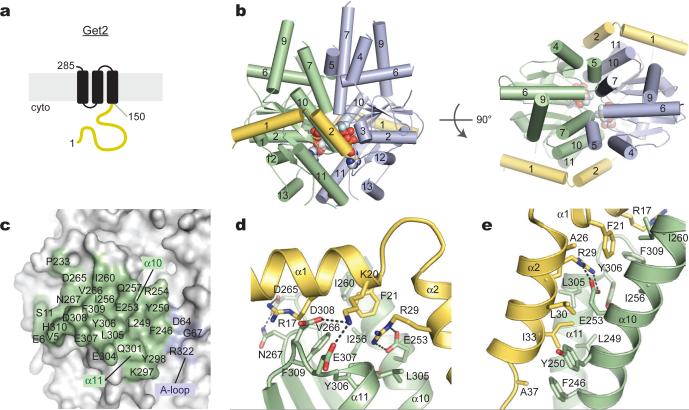
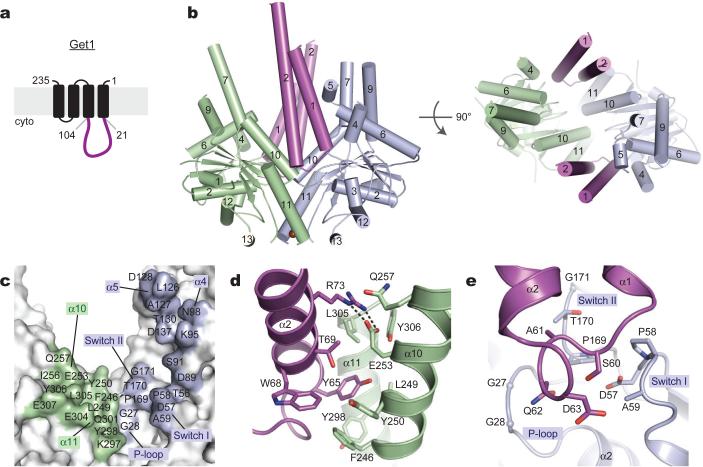
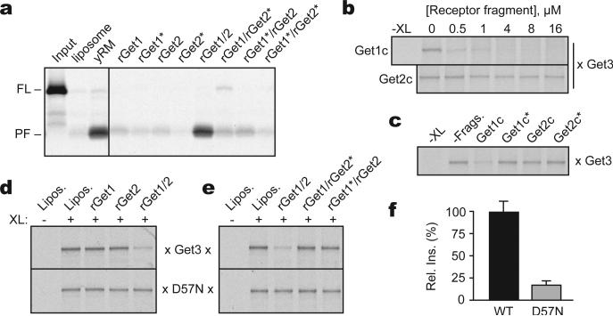
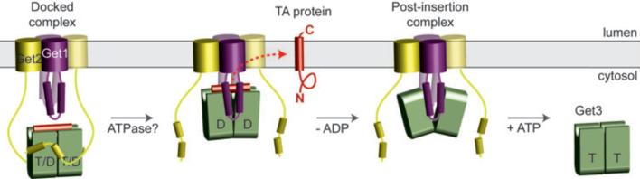
Tail-anchored (TA) membrane proteins destined for the endoplasmic reticulum are chaperoned by cytosolic targeting factors that deliver them to a membrane receptor for insertion. Although a basic framework for TA protein recognition is now emerging, the decisive targeting and membrane insertion steps are not understood. Here we reconstitute the TA protein insertion cycle with purified components, present crystal structures of key complexes between these components and perform mutational analyses based on the structures. We show that a committed targeting complex, formed by a TA protein bound to the chaperone ATPase Get3, is initially recruited to the membrane through an interaction with Get2. Once the targeting complex has been recruited, Get1 interacts with Get3 to drive TA protein release in an ATPase-dependent reaction. After releasing its TA protein cargo, the now-vacant Get3 recycles back to the cytosol concomitant with ATP binding. This work provides a detailed structural and mechanistic framework for the minimal TA protein insertion cycle.


Figures

Figure 1
Figure 1. Reconstitution of TA protein insertion with purified components
(a) Yeast microsomes (yRM) from the indicated strains were tested for insertion of purified Get3-Sec61β targeting complex (top) or by immunoblotting (bottom). The protease-protected fragment (PF) is diagnostic of successful insertion. Liposomes are a negative control. (b) Quantification of Get1 and Get2 levels in yRM by immunoblotting. (c) Protein composition of yRM and proteoliposomes containing recombinant proteins. Twenty-fold relative excess of proteoliposomes were analyzed. (d) Insertion of purified targeting complexes into liposomes, yRM, or rGet1/2 proteoliposomes. VAMP2 and Sed5 indicate TMDs from rat VAMP2 or yeast Sed5. Get1/2 concentration is indicated. (e) Insertion of purified Get3-Sec61β targeting complex into different rGet1/2 proteoliposomes. Autoradiographs and quantified data are shown.
Figure 2
Figure 2. The Get2 fragment complex with ADP•AlF4--bound Get3
(a) Predicted topology of S. cerevisiae Get2 with large cytosolic-facing region (yellow). (b) Structure of two Get2 fragments (yellow) bound to the closed Get3 dimer (green, blue). Two Mg2+ADP•AlF4- complexes and a zinc atom are indicated (spheres). An orthogonal view into the substrate-binding composite hydrophobic groove is shown on the right. (c) Get3 residues in the Get2 interface are indicated; most contacts are to one Get3 monomer (green). (d) Closeup of interactions along helix α1 of Get2, including R17, K20 and F21. (e) Closeup of interactions along helix α2 of Get2, including the conserved salt bridge between R29 and E253.
Figure 3
Figure 3. The Get1 fragment complex with Get3
(a) Predicted topology of S. cerevisiae Get1 with large cytosolic-facing region (magenta). (b) Structure of two Get1 fragments (magenta) bound to the open dimer state of Get3 (green, blue). The composite hydrophobic groove is completely disrupted. (c) Get3 residues in the Get1 interface are indicated; significant contacts are made to both monomers (green, blue). (d) Closeup of interactions between Get1 helix α2 (magenta) and one Get3 monomer (green), including the conserved salt bridge between R73 and E253. This interface overlaps extensively with the Get2c binding surface (see Fig. 2e and S11). (e) Closeup of interactions between the Get1 hairpin loop and the active site of the adjacent Get3 monomer (blue).
Figure 4
Figure 4. Mutational analysis of Get1/2/3 function
(a) Insertion assay with purified Get3-Sec61β targeting complex and proteoliposomes containing the indicated purified proteins. Liposomes and yRM are controls. Get1* and Get2* indicate mutants inactive in Get3 interaction (R73E and R17E, respectively). (b) Substrate-release from targeting complexes incubated with Get1c or Get2c; release was monitored by loss of the crosslink between radiolabeled substrate and Get3. (c) As in (b), with wild-type and mutant fragments at 0.5 μM. (d) Substrate interaction with Get3 or the ATPase-deficient Get3(D57N) was assessed by crosslinking after incubation with liposomes or proteoliposomes containing the indicated recombinant proteins. (e) As in (d), but comparing wild-type and mutant Get1/2 complexes. (f) Relative insertion efficiency into rGet1/2 proteoliposomes of targeting complexes prepared with wild-type Get3 or Get3(D57N).
Figure 5
Figure 5. ATP-dependent recycling of empty Get3 from Get1
(a) Insertion of purified Get3-Sec61β targeting complex into the indicated vesicles with or without an ATP regenerating system. (b) Proteoliposomes containing rGet1/2, or rGet1/2 bound with Get3 (left panel), were tested for insertion activity of purified targeting complex in the presence or absence of ATP (right panel). (c) Purified targeting complex was tested for insertion into wild-type yRMs or those from a ΔGet3 strain, with or without ATP. (d) Closeup of the Get1c-Get3 complex (magenta and blue) modeled onto the active site of the closed, ADP•AlF4--bound Get3 dimer (grey). Steric (dashed lines) and electrostatic clashes between conserved residues in Get1 and the nucleotide $-phosphate are apparent. (e) Dissociation of Get3-Get1c, monitored by FRET, upon titration with the indicated nucleotides. Curve fits of triplicate measurements (mean +/- s.e.m.) are shown. The reaction contained 10 nM Get3(D57N) and 100 nM Get1c. (f) As in (e), but with 10 nM Get3(D57N) and 200 nM Get2c.
Figure 6
Figure 6. Model for TA protein insertion
Nucleotide- and TA substrate-bound Get3 in a closed-dimer conformation forms the ‘docked complex’ by association with Get2. Following ATP hydrolysis, Get1 interacts with and orients Get3 along the membrane surface. This stabilizes the open-dimer conformation of Get3, disrupts the composite hydrophobic groove, and promotes TA substrate release for membrane insertion. The Get3-Get1 ‘post-insertion complex’ is dissociated by ATP binding, recycling Get3 back to the cytosol. See Supplementary Discussion for more details.

Comment in

  • It takes two to Get3.
    Sinning I, Bange G, Wild K. Sinning I, et al. Structure. 2011 Oct 12;19(10):1353-5. doi: 10.1016/j.str.2011.10.001. Structure. 2011. PMID: 22000508

References

    1. Kutay U, Hartmann E, Rapoport TA. A class of membrane proteins with a C-terminal anchor. Trends Cell Biol. 1993;3:72–75. - PubMed
    1. Beilharz T, Egan B, Silver PA, Hofmann K, Lithgow T. Bipartite signals mediate subcellular targeting of tail-anchored membrane proteins in Saccharomyces cerevisiae. J Biol Chem. 2003;278:8219–8223. - PubMed
    1. Kalbfleisch T, Cambon A, Wattenberg BW. A bioinformatics approach to identifying tail-anchored proteins in the human genome. Traffic. 2007;8:1687–1694. - PubMed
    1. Kriechbaumer V, et al. Subcellular distribution of tail-anchored proteins in Arabidopsis. Traffic. 2009;10:1753–1764. - PubMed
    1. Favaloro V, Spasic M, Schwappach B, Dobberstein B. Distinct targeting pathways for the membrane insertion of tail-anchored (TA) proteins. J Cell Sci. 2008;121:1832–1840. - PMC - PubMed

Publication types

MeSH terms

Substances