. 2011 Aug 24;476(7361):409-13.
doi: 10.1038/nature10346.

Germ-layer and lineage-restricted stem/progenitors regenerate the mouse digit tip

Affiliations

Germ-layer and lineage-restricted stem/progenitors regenerate the mouse digit tip

Yuval Rinkevich et al. Nature. .

Abstract

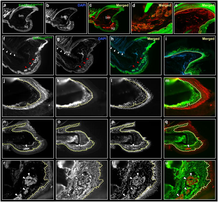
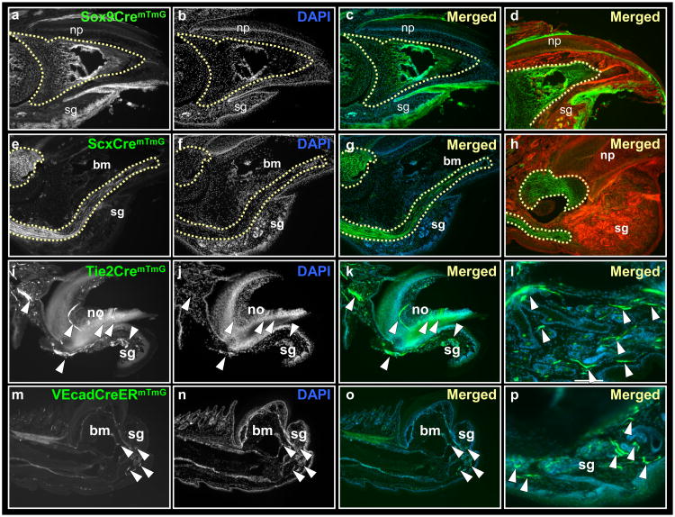
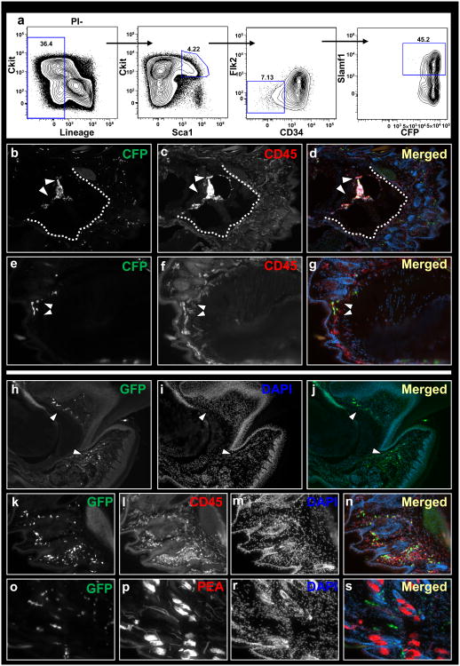
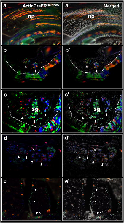
The regrowth of amputated limbs and the distal tips of digits represent models of tissue regeneration in amphibians, fish and mice. For decades it had been assumed that limb regeneration derived from the blastema, an undifferentiated pluripotent cell population thought to be derived from mature cells via dedifferentiation. Here we show that a wide range of tissue stem/progenitor cells contribute towards the restoration of the mouse distal digit. Genetic fate mapping and clonal analysis of individual cells revealed that these stem cells are lineage restricted, mimicking digit growth during development. Transplantation of cyan-fluorescent-protein-expressing haematopoietic stem cells, and parabiosis between genetically marked mice, confirmed that the stem/progenitor cells are tissue resident, including the cells involved in angiogenesis. These results, combined with those from appendage regeneration in other vertebrate subphyla, collectively demonstrate that tissue stem cells rather than pluripotent blastema cells are an evolutionarily conserved cellular mode for limb regeneration after amputation.


Figures

Figure 1
Figure 1
Germ layer restriction of ectoderm/mesoderm during digit tip regeneration. Sections through a distal digit of K14CreERmTmG (a-e), En1CremTmG (f-i) and Prx1CremTmG (j-u) transgenic mice, following three months post-amputation. Ectoderm contributes to epidermis, nail and sweat glands and fails to contribute to mesoderm tissues (a-i). Dashed line outlines the border between epidermis/dermis (d) and nail plate/matrix (e, i). Segregation of ectoderm in En1CremTmG into dorsal and ventral fates; ventral ectoderm contributes to ventral epidermis and sweat glands (f-h, red arrowheads) with no contributions to the dorsal epidermis or hair follicles (f-h, white arrowheads). A partial contribution to nail reveals dorsal and ventral chimeric origins to the nail plate (i). This boundary is shifted compared to the published lineage mapping study, using En1CreER. Lineage tracing of Prx1Cre shows restricted GFP expression to bone, tendon and mesenchyme, with no contribution to ectoderm. Keratin-14 (K14) expression in Prx1CremTmG digits is mutually exclusive from GFP expression. High magnifications of nail (j-m), bone (n-q) and sweat glands (r-u). Dashed lines outline borders between nail plate/matrix (j-m) and epidermis/dermis (n-u). White arrowheads (r-u) shows sweat glands within ventral mesenchyme are GFP negative. bm, bone marrow; sg, sweat glands.
Figure 2
Figure 2
Lineage restriction of bone, tendons and endothelium during digit tip regeneration. Sections through Sox9CremTmG (a-d), ScxCremTmG (e-h), Tie2CremTmG (i-l) and VEcadherinCreERmTmG (m-p) transgenic mice, following three months post amputations. Lineage tracing of Sox9 shows expression within all epidermal lineages. Within mesoderm, GFP expression is restricted to the distal digit bone (a-d, outlined by a dashed line). Lineage tracing of Scleraxis shows restriction of GFP to tendons (e-h, outlined by a dashed line). Lineage tracing of Tie2 and VEcadherin shows restriction of GFP in blood cells and blood vessels of the distal digit (i-p, white arrowheads). bm, bone marrow; np, nail plate, no, nail organ; sg, sweat glands.
Figure 3
Figure 3
Circulating cells do not contribute to regenerating tissues of the digit tip. Flow chart showing gating of HSCs on the basis of Lin (CD3 CD4 CD8 Mac Gr-1 B220 Ter119) Flk2 CD34 Sca+ Slamf1+ surface expressions (a). Sections through the digit of a host mice that was infused with HSCs following three months post-amputation. HSC-derived cells within bone marrow (b-d) and dermis (e-g). Dotted line outlines bone/epidermis border. Parabiosis between wild-type and genetically marked (GFP) littermates (h-r). Circulating cells within nail organ, mesenchyme surrounding sweat glands and dermis of regenerated digits (h-j, white arrowheads). Circulating cells within the regenerated digit express the hematopoietic marker CD45 (k-n) but not the endothelial marker PEA (o-r).
Figure 4
Figure 4
Multiple lineage-restricted clones contribute to digit tip regeneration. Sections through regenerated digits of ActinCreERRainbow mice. Expanding clones within nail (a-a′), epidermis (b-b′) and sweat glands (c-c′). Dashed line (b & c) outlines the epidermis/dermis border; white arrowheads show clones within ventral sweat glands. Sweat glands (d-d′) and the surrounding blood vessels (d-d′, white arrowheads) are derived from separate clones. Expanding clones within the digit bone (e-e′, white arrowheads). np, nail plate, sg, sweat glands.

References

    1. Rinkevich B, Shlemberg Z, Fishelson L. Whole-body protochordate regeneration from totipotent blood cells. Proc Natl Acad Sci U S A. 1995;92:7695–7699. - PMC - PubMed
    1. Rinkevich Y, Paz G, Rinkevich B, Reshef R. Systemic bud induction and retinoic acid signaling underlie whole body regeneration in the urochordate Botrylloides leachi. PLoS Biol. 2007;5:e71. - PMC - PubMed
    1. Voskoboynik A, et al. Striving for normality: whole body regeneration through a series of abnormal generations. FASEB J. 2007;21:1335–44. - PubMed
    1. Sánchez Alvarado A. Regeneration in the metazoans: why does it happen? Bioessays. 2000;22:578–590. - PubMed
    1. Han M, Yang X, Lee J, Allan CH, Muneoka K. Development and regeneration of the neonatal digit tip in mice. Dev Biol. 2008;315:125–135. - PMC - PubMed

Publication types

MeSH terms