Alterations of epithelial stem cell marker patterns in human diabetic corneas and effects of c-met gene therapy
- PMID: 21866211
- PMCID: PMC3159681
Alterations of epithelial stem cell marker patterns in human diabetic corneas and effects of c-met gene therapy
Abstract
Purpose: We have previously identified specific epithelial proteins with altered expression in human diabetic central corneas. Decreased hepatocyte growth factor receptor (c-met) and increased proteinases were functionally implicated in the changes of these proteins in diabetes. The present study examined whether limbal stem cell marker patterns were altered in diabetic corneas and whether c-met gene overexpression could normalize these patterns.
Methods: Cryostat sections of 28 ex vivo and 26 organ-cultured autopsy human normal and diabetic corneas were examined by immunohistochemistry using antibodies to putative limbal stem cell markers including ATP-binding cassette sub-family G member 2 (ABCG2), N-cadherin, ΔNp63α, tenascin-C, laminin γ3 chain, keratins (K) K15, K17, K19, β(1) integrin, vimentin, frizzled 7, and fibronectin. Organ-cultured diabetic corneas were studied upon transduction with adenovirus harboring c-met gene.
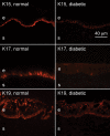
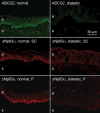
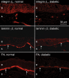
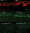
Results: Immunostaining for ABCG2, N-cadherin, ΔNp63α, K15, K17, K19, and β(1) integrin, was significantly decreased in the stem cell-harboring diabetic limbal basal epithelium either by intensity or the number of positive cells. Basement membrane components, laminin γ3 chain, and fibronectin (but not tenascin-C) also showed a significant reduction in the ex vivo diabetic limbus. c-Met gene transduction, which normalizes diabetic marker expression and epithelial wound healing, was accompanied by increased limbal epithelial staining for K17, K19, ΔNp63α, and a diabetic marker α(3)β(1) integrin, compared to vector-transduced corneas.
Conclusions: The data suggest that limbal stem cell compartment is altered in long-term diabetes. Gene therapy, such as with c-met overexpression, could be able to restore normal function to diabetic corneal epithelial stem cells.
Figures


References
-
- Herse PR. A review of manifestations of diabetes mellitus in the anterior eye and cornea. Am J Optom Physiol Opt. 1988;65:224–30. - PubMed
-
- Cavallerano J. Ocular manifestations of diabetes mellitus. Optom Clin. 1992;2:93–116. - PubMed
-
- Saini JS, Khandalavla B. Corneal epithelial fragility in diabetes mellitus. Can J Ophthalmol. 1995;30:142–6. - PubMed
-
- Sánchez-Thorin JC. The cornea in diabetes mellitus. Int Ophthalmol Clin. 1998;38:19–36. - PubMed
-
- Malik RA, Kallinikos P, Abbott CA, van Schie CH, Morgan P, Efron N, Boulton AJ. Corneal confocal microscopy: a non-invasive surrogate of nerve fibre damage and repair in diabetic patients. Diabetologia. 2003;46:683–8. - PubMed
Publication types
MeSH terms
Substances
Grants and funding
LinkOut - more resources
Full Text Sources
Medical
Research Materials
Miscellaneous
