. 2010 Jun 1:2:9-16.
doi: 10.2147/OAAP.S7202.

Auditory sensitivity and the outer hair cell system in the CBA mouse model of age-related hearing loss

Affiliations

Auditory sensitivity and the outer hair cell system in the CBA mouse model of age-related hearing loss

Robert D Frisina et al. Open Access Anim Physiol. .

Abstract

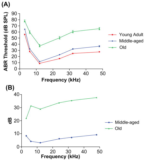
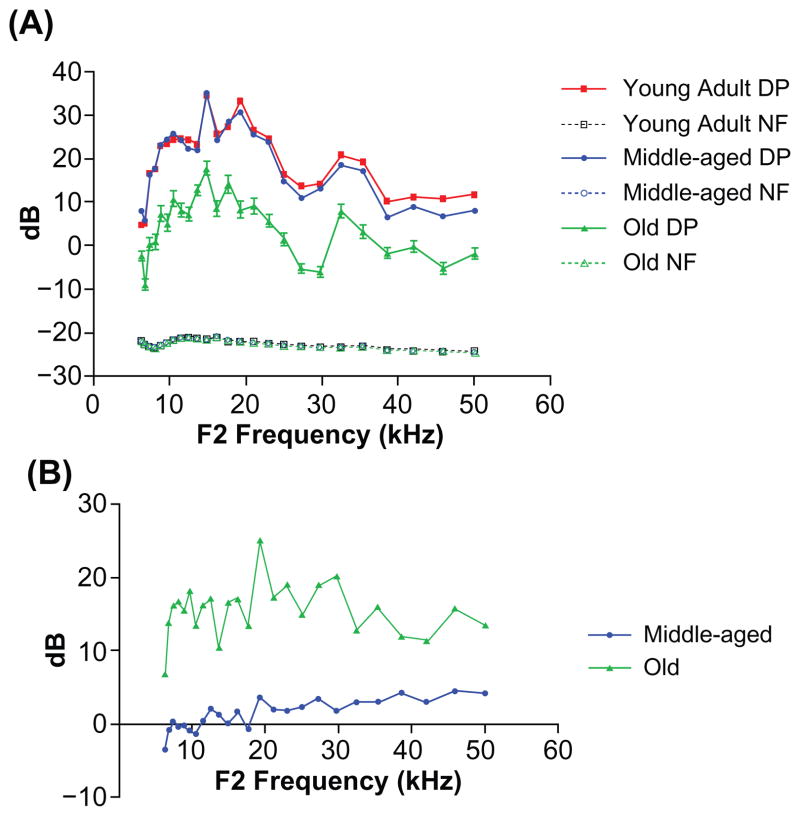
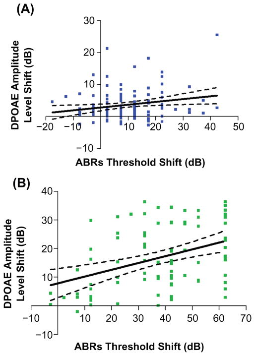
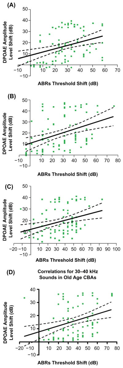
Age-related hearing loss is a highly prevalent sensory disorder, from both the clinical and animal model perspectives. Understanding of the neurophysiologic, structural, and molecular biologic bases of age-related hearing loss will facilitate development of biomedical therapeutic interventions to prevent, slow, or reverse its progression. Thus, increased understanding of relationships between aging of the cochlear (auditory portion of the inner ear) hair cell system and decline in overall hearing ability is necessary. The goal of the present investigation was to test the hypothesis that there would be correlations between physiologic measures of outer hair cell function (otoacoustic emission levels) and hearing sensitivity (auditory brainstem response thresholds), starting in middle age. For the CBA mouse, a useful animal model of age-related hearing loss, it was found that correlations between these two hearing measures occurred only for high sound frequencies in middle age. However, in old age, a correlation was observed across the entire mouse range of hearing. These findings have implications for improved early detection of progression of age-related hearing loss in middle-aged mammals, including mice and humans, and distinguishing peripheral etiologies from central auditory system decline.


Conflict of interest statement

Disclosures

The authors report no conflicts of interest in this work.

Figures

Figure 1
Figure 1
Comparisons of ABR thresholds in three mouse age groups. A) Frequency threshold curves for the young adult and middle ages were similar, but a large threshold elevation occurred in old age. The variability of the responses showed a marked increase in the old group, as evidenced by the larger error bars. B) ABR threshold shift comparisons between young adult CBA mice and the two older age groups. The middle-aged mice had thresholds within about 10 dB of the young adults. The old group had 22–38 dB ABR threshold elevations compared with young adults. Both of the older subject groups displayed a high frequency hearing loss. Abbreviation: ABR auditory, brainstem response.
Figure 2
Figure 2
The DPOAE-gram sreveal significant differences between the old subjects and the other two age groups. A) DPOAE levels were similar between the young adults and middle-aged mice, but large declines occurred in old age. Note the larger error bars in the oldest group. B) DPOAE level shifts compared with young adult: the middle-aged subjects showed a 2–5 dB change in the middle and high frequencies of the mouse hearing range, but there were large 10–25 dB shifts for the old group. Abbreviations: DP, distortion product; DPOAE, distortion-product otoacoustic emission; NF, noise floor.
Figure 3
Figure 3
Group correlations between decline in the outer hair cell system and overall auditory sensitivity start to occur in middle age. A) Correlations between ABR thresholds and DPOAE level shifts are significant, only at the higher frequencies (30–50 kHz) in middle age: F(1,124) = 5.32, P = 0.0228, r2 = 0.0411. B) The correlations in the high frequency range become much stronger in old age: F (1,84) =15.21, P = 0.0002, r2 = 0.1533. Here, and in the remaining figures, dashed lines in the regression plots represent 95% confidence intervals. Abbreviations: ABR, auditory brainstem response; DPOAE, distortion-product otoacoustic emission.
Figure 4
Figure 4
In old age, regression analyses yielded significant relationships between otoacoustic emission levels and ABR thresholds for all of the frequency bands tested. A) 1–10 kHz, F (1,84) = 12.90, P = 0.0006, r2 = 0.1331. B) 10–20 kHz, F (1,84) = 14.64, P = 0.0002, r2 = 0.1484. C) 20–30 kHz, F (1,84) = 10.08, P = 0.0021, r2 = 0.1071. D) 30–40 kHz, F (1,84) =10.70, P = 0.0016, r2 = 0.1130. Abbreviation: ABR, auditory brainstem response.
Figure 5
Figure 5
Correlations between ABR thresholds and DPOAE levels across the entire hearing range for mice of different ages. A) Correlations for the middle age mice were present, but not as striking as in old age: F(1,124) =4.22, P = 0.042, r2 = 0.033. B) A greater relationship was observed in the old mice: F(1,84) =18.43, P < 0.0001, r2 = 0.18. Note: Since young adults all had excellent hearing, there were no significant relationships. Abbreviations: ABR, auditory brain stem response; DPOAE, distortion-product otoacoustic emission.

References

    1. Schmiedt RA, Lang H, Okamura H, Schulte BA. Effects of furosemide applied chronically to the round window: A model of metabolic presbycusis. J Neurosci. 2002;22:9643–9650. - PMC - PubMed
    1. Lang H, Schulte BA, Schmiedt RA. Effects of chronic furosemide treatment and age on cell division in the adult gerbil inner ear. J Assoc Res Otolaryngol. 2003;4:164–175. - PMC - PubMed
    1. Casey MA, Feldman ML. Aging in the rat medial nucleus of the trapezoid body. I. Light microscopy. Neurobiol Aging. 1982;3:187–195. - PubMed
    1. Caspary DM, Milbrandt JC, Helfert RH. Central auditory aging: GABA changes in the inferior colliculus. Exp Gerontol. 1995;30:349–360. - PubMed
    1. Caspary DM, Raza A, Lawhorn Armour BA, Pippin J, Arneric SP. Immunocytochemical and neurochemical evidence for age-related loss of GABA in the inferior colliculus: Implications for neural presbycusis. J Neurosci. 1990;10:2363–2372. - PMC - PubMed

LinkOut - more resources