. 2011 Aug 1:5:64.
doi: 10.3389/fnsys.2011.00064. eCollection 2011.

The corticostriatal and corticosubthalamic pathways: two entries, one target. So what?

Affiliations

The corticostriatal and corticosubthalamic pathways: two entries, one target. So what?

Abraham Mathai et al. Front Syst Neurosci. .

Abstract

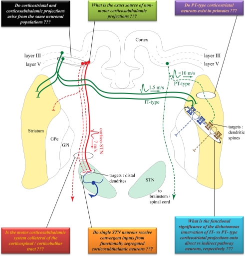
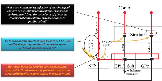
The basal ganglia receive cortical inputs through two main stations - the striatum and the subthalamic nucleus (STN). The information flowing along the corticostriatal system is transmitted to the basal ganglia circuitry via the "direct and indirect" striatofugal pathways, while information that flows through the STN is transmitted along the so-called "hyperdirect" pathway. The functional significance of this dual entry system is not clear. Although the corticostriatal system has been thoroughly characterized anatomically and electrophysiologically, such is not the case for the corticosubthalamic system. In order to provide further insights into the intricacy of this complex anatomical organization, this review examines and compares the anatomical and functional organization of the corticostriatal and corticosubthalamic systems, and highlights some key issues that must be addressed to better understand the mechanisms by which these two neural systems may interact to regulate basal ganglia functions and dysfunctions.

Keywords: Parkinson's disease; basal ganglia; cerebral cortex; deep brain stimulation; hyperdirect; monkey; striatum; subthalamic nucleus.


Figures

Figure 1
Figure 1
Comparison of the cortical sources, conduction velocities and synaptic targets of corticostriatal versus corticosubthalamic projections. Solid lines indicate well established projections. Dotted lines indicate projections that need to be confirmed in future studies. Colored boxes summarize some of the unanswered questions pertaining to the anatomical, synaptic, and functional organization of these two pathways. See text for abbreviations. Anatomical structures are not to scale, but depicted solely for illustrative purposes and are not necessarily in the same coronal plane. Some connections have been purposefully omitted to decrease complexity.
Figure 2
Figure 2
Highly simplified overview of the classical model of the basal ganglia circuitry. Red lines indicate excitatory projections. Black lines indicate inhibitory projections. Some brainstem connections have been omitted for the sake of simplicity. Colored boxes highlight some key issues to be addressed about the anatomy and function of the corticostriatal and corticosubthalamic systems. See text for abbreviations.
Figure 3
Figure 3
Functional domains of the STN. Coronal view of the primate STN to illustrate the topographical organization of cortical afferents to various functional regions of the nucleus defined by their reciprocal connections with the globus pallidus and motor cortical areas. Solid lines indicate well established corticosubthalamic projections from motor and pre-motor cortices. Dotted lines indicate putative projections that need further confirmation. Abbreviations: cing, cingulate.

References

    1. Afsharpour S. (1985). Topographical projections of the cerebral cortex to the subthalamic nucleus. J. Comp. Neurol. 236, 14–2810.1002/cne.902360103 - DOI - PubMed
    1. Albin R. L., Young A. B., Penney J. B. (1989). The functional anatomy of basal ganglia disorders. Trends Neurosci. 12, 366–37510.1016/0166-2236(89)90074-X - DOI - PubMed
    1. Alexander G. E., Delong M. R., Strick P. L. (1986). Parallel organization of functionally segregated circuits linking basal ganglia and cortex. Annu. Rev. Neurosci. 9, 357–38110.1146/annurev.ne.09.030186.002041 - DOI - PubMed
    1. Arbuthnott G. W., Ingham C. A., Wickens J. R. (2000). Dopamine and synaptic plasticity in the neostriatum. J. Anat. 196(Pt 4), 587–59610.1046/j.1469-7580.2000.19640499.x - DOI - PMC - PubMed
    1. Aron A. R., Behrens T. E., Smith S., Frank M. J., Poldrack R. A. (2007). Triangulating a cognitive control network using diffusion-weighted magnetic resonance imaging (MRI) and functional MRI. J. Neurosci. 27, 3743–375210.1523/JNEUROSCI.0519-07.2007 - DOI - PMC - PubMed

LinkOut - more resources