. 2012 Jan;113(1):49-60.
doi: 10.1002/jcb.23326.

Inhibition of Wnt signaling by cucurbitacin B in breast cancer cells: reduction of Wnt-associated proteins and reduced translocation of galectin-3-mediated β-catenin to the nucleus

Affiliations

Inhibition of Wnt signaling by cucurbitacin B in breast cancer cells: reduction of Wnt-associated proteins and reduced translocation of galectin-3-mediated β-catenin to the nucleus

Sumana Dakeng et al. J Cell Biochem. 2012 Jan.

Abstract

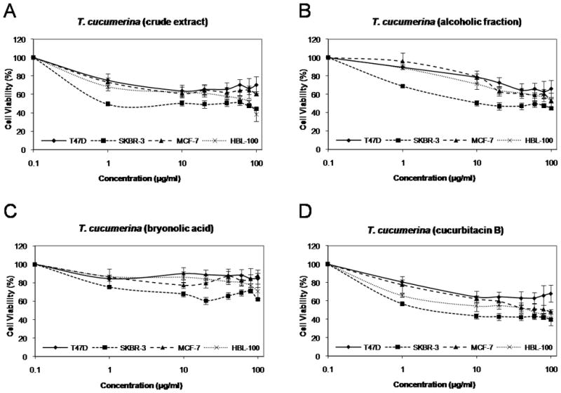
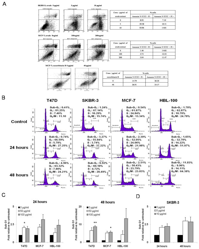
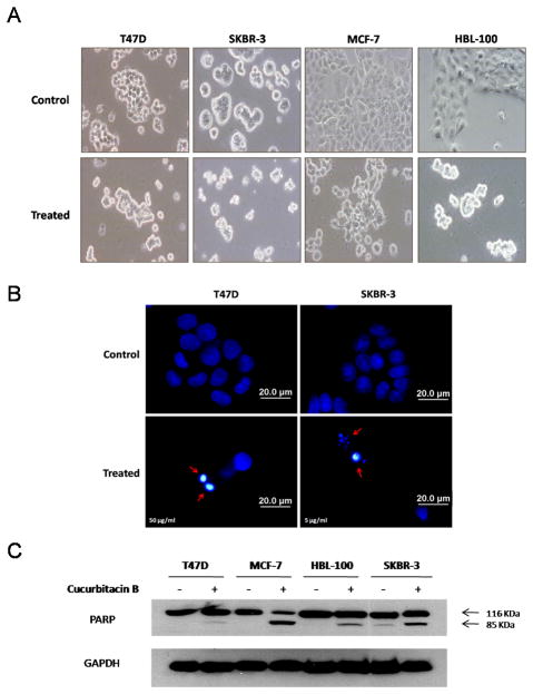
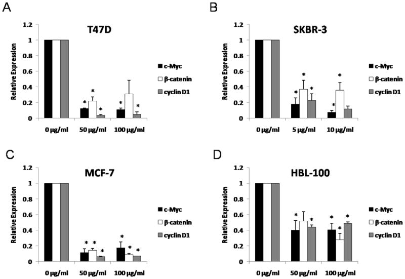
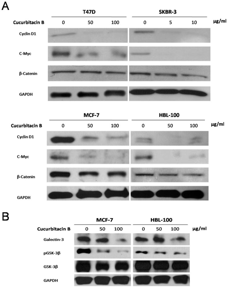
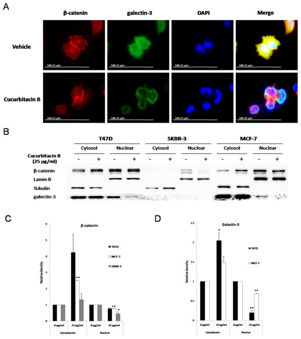
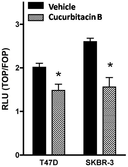
The cucurbitacins are tetracyclic triterpenes found in plants of the family Cucurbitaceae. Cucurbitacins have been shown to have anti-cancer and anti-inflamatory activities. We investigated the anti-cancer activity of cucurbitacin B extracted from Thai medicinal plant Trichosanthes cucumerina Linn. Cell viability was assessed by MTT (3-(4,5-dimethylthiazol-2-yl)-2,5-diphenyltetrazolium bromide) assay. Results indicated that cucurbitacin B from T. cucumerina Linn. has a cytotoxic effect on breast cancer cell lines SKBR-3 and MCF-7 with an IC50 of 4.60 and 88.75 µg/ml, respectively. Growth inhibition was attributed to G2/M phase arrest and apoptosis. Cyclin D1, c-Myc, and β-catenin expression levels were reduced. Western blot analysis showed increased PARP cleavage and decreased Wnt-associated signaling molecules β-catenin, galectin-3, cyclin D1 and c-Myc, and corresponding changes in phosphorylated GSK-3β levels. Cucurbitacin B treatment inhibited translocation to the nucleus of β-catenin and galectin-3. The depletion of β-catenin and galectin-3 in the nucleus was confirmed by cellular protein fractionation. T-cell factor (TCF)/lymphoid enhancer factor (LEF)-dependent transcriptional activity was disrupted in cucurbitacin B treated cells as tested by a TCF reporter assay. The relative luciferase activity was reduced when we treated cells with cucurbitacin B compound for 24 h. Our data suggest that cucurbitacin B may in part induce apoptosis and exert growth inhibitory effect via interruption the Wnt signaling.


Figures

Figure 1
Figure 1
Effect of T. cucumerina extracts on the growth of breast cancer cells. Cell number was measured using the MTT assay after treatment with various T. cucumerina extracts; A: crude spray-dried extract, B: alcoholic fraction, C: bryonolic acid fraction or D: cucurbitacin B compound.
Figure 2
Figure 2
Cucurbitacin B promotes G2/M phase arrest and induces apoptosis. A: Percent positive cells after staining with Annexin V-FITC and PI after SKBR-3 and MCF-7 were treated with crude spray-dried T. cucumerina Linn. or cucurbitacin B for 24 hours. Percentage of Annexin V-FITC (+)/ PI (−) cells indicates the early apoptotic fraction and Annexin V-FITC (+)/ PI (+) indicates cells in late apoptosis. B: Cell cycle analysis was performed in T47D, SKBR-3, MCF-7 and HBL-100 cell lines treated with cucurbitacin B at half maximal concentration (IC50). Cell cycle/DNA content was analyzed by flow cytometry after 24 and 48 hours. The percentages of cells at each cell cycle phase are shown in each panel. C: Sub G1 population relative to untreated cells was measured when treated T47D, MCF-7 and HBL-100 with 50 and 100 µg/ml. D: SKBR-3 with 5 and 10 µg/ml of cucurbitacin B (10-times less concentration) for 24 and 48 hours. * P< 0.05, ** P< 0.01 compared with the untreated control.
Figure 3
Figure 3
Morphological changes, apoptotic bodies, nuclear fragmentation and PARP cleavage induced by cucurbitacin B. Cells were treated with 50 µg/ml (T47D, MCF-7, HBL-100) and 5 µg/ml (SKBR-3) for 24 hours. A: Phase-contrast photomicrographs of untreated control (upper panel) and treated cells (lower panel). B: Cucurbitacin B treated cells were stained with DAPI to differentiate non-apoptotic from apoptotic cells (arrows). Staining was analyzed by fluorescence microscopy. C: Cell lysates were analyzed by Western blotting using anti-PARP which detects both intact PARP (116 kDa) and apoptotic marker PARP cleavage fragment (85 kDa). GAPDH was used as the loading control.
Figure 4
Figure 4
Expressions of c-Myc, β-catenin and cyclin D1 after cucurbitacin B treatment. A: T47D, B: SKBR-3, C: MCF-7 and D: HBL-100 cells were incubated with the specified concentrations of cucurbitacin B for 24 hours and the quantitative expression levels of c-Myc, β-catenin, and cyclin D1 were analyzed by real-time RT-PCR. Results shown are the average of three independent experiments. * P< 0.05 compared with the control group.
Figure 5
Figure 5
Differential expression of proteins associated with Wnt signaling. Western blot analysis was performed to compare expression levels of A: cyclin D1, c-Myc and β-catenin or B: galectin-3, GSK-3β and phosphorylated GSK-3β proteins among cucurbitacin B treated and untreated cells. Expression of GAPDH was used as protein loading control.
Figure 6
Figure 6
Nuclear translocation of β-catenin and galectin-3 was inhibited by cucurbitacin B. A: T47D cells were treated either with 25 µg/ml cucurbitacin B or control medium for 24 hours, fixed and immunostained with anti-β-catenin and anti-galectin-3 antibodies. Immunostaining was analyzed using fluorescence microscope. B: Three breast cancer cells T47D, SKBR-3 and MCF-7 were treated with 25 µg/ml cucurbitacin B for 24 hours. Cellular fractionation was carried out to determine the cellular localization of β-catenin and galectin-3. Lamin B was used as loading control for nuclear fraction and tubulin as control for cytoplasmic fraction. C: Relative density of cytoplasmic and nuclear proteins was analyzed. Results shown are the average of three independent experiments. * P< 0.05, ** P< 0.01 compared with the vehicle control.
Figure 7
Figure 7
TCF reporter activity was decreased in cucurbitacin B treated cells. T47D and SKBR-3 were transiently transfected with either TOPFLASH or FOPFLASH together with renilla (as control expression). Cells were then treated with 25 µg/ml cucurbitacin B or vehicle control medium for 24 hours. Luciferase assay was measured and results were expressed as the ratio of TOPFLASH over FOPFLASH activity. The significance of the difference in the repression of TOPFLASH by vehicle and cucurbitacin B was analyzed using a paired Student’s t test. * P< 0.05 compared with the vehicle control.

References

    1. Arawwawala M, Thabrew I, Arambewela L, Handunnetti S. Anti-inflammatory activity of Trichosanthes cucumerina Linn. in rats. J Ethnopharmacol. 2010;131:538–543. - PubMed
    1. Blagosklonny MV, Fojo T. Molecular effects of paclitaxel: myths and reality (a critical review) Int J Cancer. 1999;83:151–156. - PubMed
    1. Bray F, Sankila R, Ferlay J, Parkin DM. Estimates of cancer incidence and mortality in Europe in 1995. Eur J Cancer. 2002;38:99–166. - PubMed
    1. Brennan KR, Brown AM. Wnt proteins in mammary development and cancer. J Mammary Gland Biol Neoplasia. 2004;9:119–131. - PubMed
    1. Brown AM. Wnt signaling in breast cancer: have we come full circle? Breast Cancer Res. 2001;3:351–355. - PMC - PubMed

Publication types

MeSH terms