

Rapidly growing left atrial myxoma: a case report
- PMID: 21867489
- PMCID: PMC3177928
- DOI: 10.1186/1752-1947-5-417
Rapidly growing left atrial myxoma: a case report
Abstract
Introduction: Left atrial myxomas are rare benign tumors of the heart. They vary widely in size, and very little is known about their growth rate. The reported growth rates of left atrial myxomas from several published case reports appears to vary from no growth, to between 1.3 to 6.9 mm/month in diameter within patients with established myxoma who have not undergone surgery.
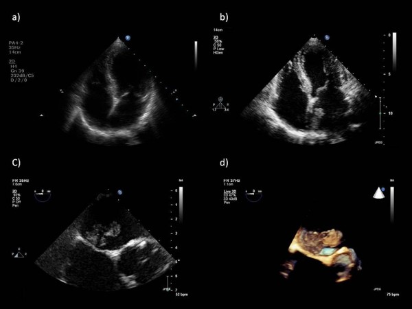
Case presentation: We present the case of a rapidly growing pedunculated left atrial myxoma in a 62-year-old asymptomatic Caucasian woman found incidentally during routine transthoracic echocardiography. Our patient was attending her annual valve clinic assessment for moderate aortic regurgitation, and her two previous consecutive transthoracic echocardiography scans performed 12 and 24 months prior to this appointment had demonstrated a clear left atrium and aortic regurgitation of moderate severity.
Conclusions: To the best of our knowledge, our case is the first to provide images of absence and presence of myxoma from transthoracic echocardiography scans taken a year apart, with estimated growth rate of 2.2 mm/month. Rapidly growing myxoma may be mistaken for thrombus, and may require urgent surgical excision to reduce the risk of associated complications such as thrombo-embolic events, sudden cardiac death and removal of a possibly malignant tumor. The potential for rapid growth should be considered if there is a plan to delay surgery. Furthermore, it would be pertinent to consider annual echocardiography in patients presenting with clinical features suggestive of cardiac myxoma such as constitutional symptoms, as these tumors may be rapid growing and may only become apparent on subsequent echocardiography.
Figures
References
LinkOut - more resources
Full Text Sources
